A groundbreaking investigation conducted by researchers at the Kennedy Institute has unveiled compelling evidence suggesting that certain joints may possess an inherent vulnerability to inflammatory arthritis even before an individual is born. This pivotal discovery offers a potential resolution to a longstanding enigma within the field of rheumatology: why rheumatoid arthritis (RA) consistently targets specific joints while largely sparing others. The findings, detailed in the prestigious journal Nature Immunology under the title "The embryonic origins of site-specific arthritis," propose a paradigm shift, indicating that the propensity for RA may not solely hinge on the immune system’s later activity, but also on intrinsic biological distinctions forged within individual joints during their very formation.
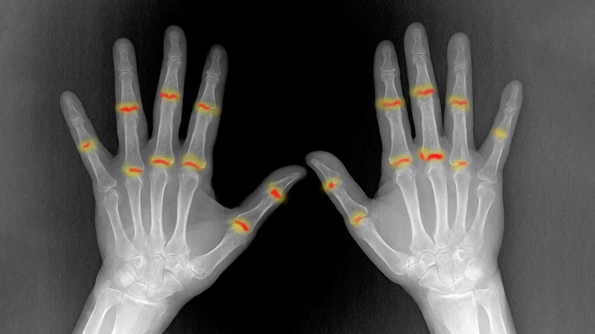
Rheumatoid arthritis stands as a debilitating autoimmune disorder characterized by the immune system erroneously attacking the synovium, the specialized connective tissue lining the inner surface of joints. This misguided assault precipitates a cascade of symptoms including profound pain, persistent swelling, and debilitating stiffness. Over time, this chronic inflammation can inflict irreversible damage upon the cartilage, underlying bone, and surrounding soft tissues, severely compromising joint function and quality of life. Affecting millions globally, RA presents a significant public health challenge, with current therapeutic strategies primarily focusing on managing symptoms and slowing disease progression through immunosuppression, yet often failing to prevent localized flare-ups or fully explain the disease’s selective anatomical targets.
For decades, clinicians and scientists have observed RA’s distinct predilection for certain anatomical sites. For instance, the proximal interphalangeal (PIP) joints – those located in the middle of the fingers – are frequently afflicted, whereas the distal interphalangeal (DIP) joints, found nearer the fingertips, are typically spared. This consistent pattern has long puzzled the medical community, prompting questions about underlying mechanisms beyond systemic immune dysregulation. The Oxford-led research team embarked on a mission to decipher this mystery, turning their investigative lens towards the earliest stages of human development.
To meticulously probe the foundational differences that might predispose joints to RA, the scientists employed a sophisticated array of advanced imaging and molecular profiling techniques. Their approach involved a comparative analysis of the PIP and DIP joints, specifically examining them during embryonic development. This strategy proved critical, as studying developing joints allowed the researchers to scrutinize entire joint structures in an unprecedented level of detail, largely unmarred by the complex immune cell infiltration and tissue remodeling characteristic of adult arthritic joints. The team leveraged single-cell sequencing, a powerful methodology that enables the analysis of gene expression at the individual cell level, thereby revealing the diverse cellular populations and their unique functions within the developing joint. Complementing this, advanced image analysis and high-resolution 3D X-ray scanning, performed at facilities such as the Diamond Light Source at the Harwell Science and Innovation Campus, provided an intricate, three-dimensional reconstruction of joint architecture and tissue organization.
The initial insights gained from this meticulous mapping of developing human finger joints revealed that these nascent structures were predominantly composed of structural cells, rather than immune cells, which are abundant in adult inflammatory conditions. Among these were various types of fibroblasts and chondrocytes, cells vital for forming cartilage. This observation underscored the importance of focusing on the intrinsic biology of these structural components. Further investigation delved into the molecular signals orchestrating the differentiation of these cells into their specialized forms, a process crucial for establishing joint integrity and function.
One particular group of cells garnered significant attention: the fibroblasts that constitute the synovial lining. These remarkable cells are indispensable for joint health, producing lubricating substances that reduce friction, protect articular surfaces, and facilitate smooth movement. However, in the context of arthritis, these same cells can undergo profound phenotypic changes, adopting an abnormal, pro-inflammatory behavior that exacerbates disease pathology. Understanding the developmental origins and regulatory pathways governing these synovial fibroblasts could unlock novel therapeutic avenues aimed at restoring their protective functions.
The research illuminated several critical differences between the RA-prone PIP joints and the relatively resistant DIP joints, even before birth. A striking observation was the significantly larger volume of synovial tissue found in PIP joints. Furthermore, using a specially developed image analysis tool, the researchers identified a higher abundance of a specific subtype of connective tissue cell, designated as PI16-positive (PI16+) fibroblasts, within the PIP joints. These PI16+ fibroblasts were strategically localized around blood vessels and at sites where tendons and ligaments connect to adjacent bone and tissue – locations often implicated in the initiation and propagation of inflammatory processes.
Dr. Sarah Davidson, a Postdoctoral Researcher at the Kennedy Institute and one of the lead authors of the study, emphasized the profound implications of these findings. "We discovered that joints commonly affected by rheumatoid arthritis already possess distinct cellular populations prenatally," Dr. Davidson stated. "PI16+ fibroblasts were enriched in these vulnerable joints and exhibited a differential response to inflammatory signals. Their specific location and behavior strongly suggest they could play a fundamental role in dictating where the disease manifests."
Further functional analysis revealed that while both PI16+ and PI16- fibroblast populations exhibited a general pro-inflammatory response when exposed to inflammatory cues, the PI16+ cells uniquely demonstrated distinct alterations in biological pathways associated with immune regulation and tissue organization. This suggests that PI16+ fibroblasts are not merely passive responders but active participants in shaping the inflammatory milieu, potentially acting as key orchestrators of the local immune response within the joint. Beyond individual cell populations, the 3D imaging data from Diamond Light Source further confirmed that the synovial tissue in PIP joints was not only more voluminous but also exhibited a distinct organizational pattern compared to the synovium in DIP joints. These multifaceted differences, encompassing both cellular composition and tissue architecture, collectively offer a compelling explanation for the differential susceptibility of various joints to inflammatory conditions like RA.
Professor Christopher Buckley, the Kennedy Professor of Translational Rheumatology at the University of Oxford and a senior author of the study, highlighted the significance of these revelations. "For many decades, the selective targeting of particular joints by rheumatoid arthritis has been one of the great unanswered questions in rheumatology," Professor Buckley remarked. "Our findings propose that the answer resides not exclusively within the immune system, but also intrinsically within the tissues themselves. The unique cellular and structural characteristics established during embryonic development appear to be critical in determining where inflammation ultimately takes hold later in life."
The study also shed light on the developmental trajectory of the synovial lining itself, suggesting that it may originate from dual sources: embryonic cartilage and surrounding joint fibroblasts. This intricate developmental process, the researchers noted, appears to be influenced by local environmental conditions, including variations in oxygen levels. Understanding these intricate developmental signals is paramount, as it could provide unprecedented clues into how synovial fibroblasts acquire their specialized functions. Such knowledge could, in turn, open pathways to novel therapeutic strategies aimed at re-establishing their normal protective behaviors and preventing their pathogenic transformation in arthritic diseases.
The implications of this research are far-reaching, potentially reshaping the fundamental understanding of rheumatoid arthritis pathogenesis. It posits a broader, more integrated explanation for the disease’s selective targeting of joints, moving beyond a purely immune-centric view. Rather than joint vulnerability being determined entirely by immune system activity in adulthood, the research suggests that the inherent tendency for inflammation to develop in certain areas is significantly influenced by cellular and structural features laid down during the very formation of the joints. In essence, each joint’s unique local biological environment, established early in life, may predetermine its susceptibility to rheumatoid arthritis years or even decades later.
This paradigm shift opens exciting new avenues for research, diagnosis, and treatment. Future investigations could focus on identifying specific biomarkers within the developing joint that predict RA susceptibility, potentially enabling earlier intervention or even preventative strategies for individuals identified as high-risk. Furthermore, understanding the distinct biological pathways and cellular interactions within PI16+ fibroblasts could lead to the development of highly targeted therapies designed to modulate their behavior, thereby mitigating inflammatory responses at their source. The collaborative effort that underpinned this research, involving scientists from the Kennedy Institute of Rheumatology at the University of Oxford, the University of Birmingham, University College London, and Diamond Light Source, underscores the interdisciplinary nature required to tackle such complex biological puzzles. Supported by the Medical Research Council (MRC), this study represents a significant stride towards a more comprehensive understanding of rheumatoid arthritis, offering hope for more effective and personalized interventions in the future.
About the Author