
Alzheimer’s disease, a pervasive neurodegenerative condition, currently impacts an estimated 7.2 million Americans aged 65 and older, according to statistics from the Alzheimer’s Association. Traditional diagnostic pathways for this debilitating illness have predominantly relied on quantifying the presence of two key proteins – amyloid-beta (Aβ) and phosphorylated tau (p-tau) – in biological fluids such as blood or cerebrospinal fluid. While these established biomarkers are instrumental in current clinical practice, there is a growing consensus among the scientific community that they may not entirely capture the earliest molecular shifts occurring within the intricate cascade of Alzheimer’s disease development.
A significant breakthrough in this diagnostic landscape has been introduced by researchers at Scripps Research, who have developed an innovative blood-based assay that diverges from conventional methods. This novel test shifts its focus from merely measuring the quantity of proteins circulating in the bloodstream to examining the intricate three-dimensional configurations – the folding patterns – of these proteins. Published on February 27, 2026, in the esteemed journal Nature Aging, the findings from this pioneering study highlight a compelling correlation between specific structural variations in three plasma proteins and an individual’s Alzheimer’s status. This sophisticated approach demonstrated a remarkable ability to differentiate between individuals with unimpaired cognitive function, those experiencing mild cognitive impairment (MCI), and patients formally diagnosed with Alzheimer’s disease. The implications of this research are profound, suggesting a future where earlier detection could pave the way for more timely and effective therapeutic interventions.
The fundamental premise underpinning this research stems from the broader understanding of neurodegenerative disorders. Professor John Yates, a senior author of the study and a distinguished professor at Scripps Research, articulates this perspective, stating, "Many neurodegenerative diseases are driven by changes in protein structure." He further elaborated on the central question that guided their investigation: "The question was, are there structural changes in specific proteins that might be useful as predictive markers?" This inquiry led them to explore the intricate relationship between protein misfolding and the progression of neurodegenerative conditions.
The Crucial Role of Proteostasis in Cellular Health
For decades, the pathological hallmarks of Alzheimer’s disease have been primarily characterized by the extracellular accumulation of amyloid plaques and the intracellular formation of tau tangles within the brain. However, contemporary scientific understanding is increasingly pointing towards a more systemic failure in a fundamental cellular process known as proteostasis. Proteostasis encompasses the complex network of mechanisms responsible for ensuring that proteins within a cell maintain their correct three-dimensional structures and for efficiently clearing out any misfolded or damaged proteins. This intricate regulatory system is vital for maintaining cellular integrity and function.
With advancing age, the efficiency of the proteostasis machinery tends to decline. This age-related decline makes proteins more susceptible to errors during their synthesis or during maintenance and repair processes, leading to an increased likelihood of adopting incorrect conformations. Building upon this understanding, the Scripps Research team hypothesized that if the proteostasis system is compromised in the brain, leading to aberrant protein structures, then similar structural anomalies might also manifest in proteins circulating throughout the peripheral bloodstream. This proposed connection between central nervous system pathology and peripheral blood markers formed the conceptual bedrock of their study.
Unraveling Protein Structural Signatures in Blood Samples
To rigorously test their hypothesis, the research team embarked on a comprehensive analysis of plasma samples obtained from a cohort of 520 participants. This carefully selected group was stratified into three distinct categories: cognitively healthy adults, individuals diagnosed with mild cognitive impairment, and patients with a confirmed diagnosis of Alzheimer’s disease. This tripartite division allowed for a nuanced examination of how protein structure might vary across different stages of cognitive decline.
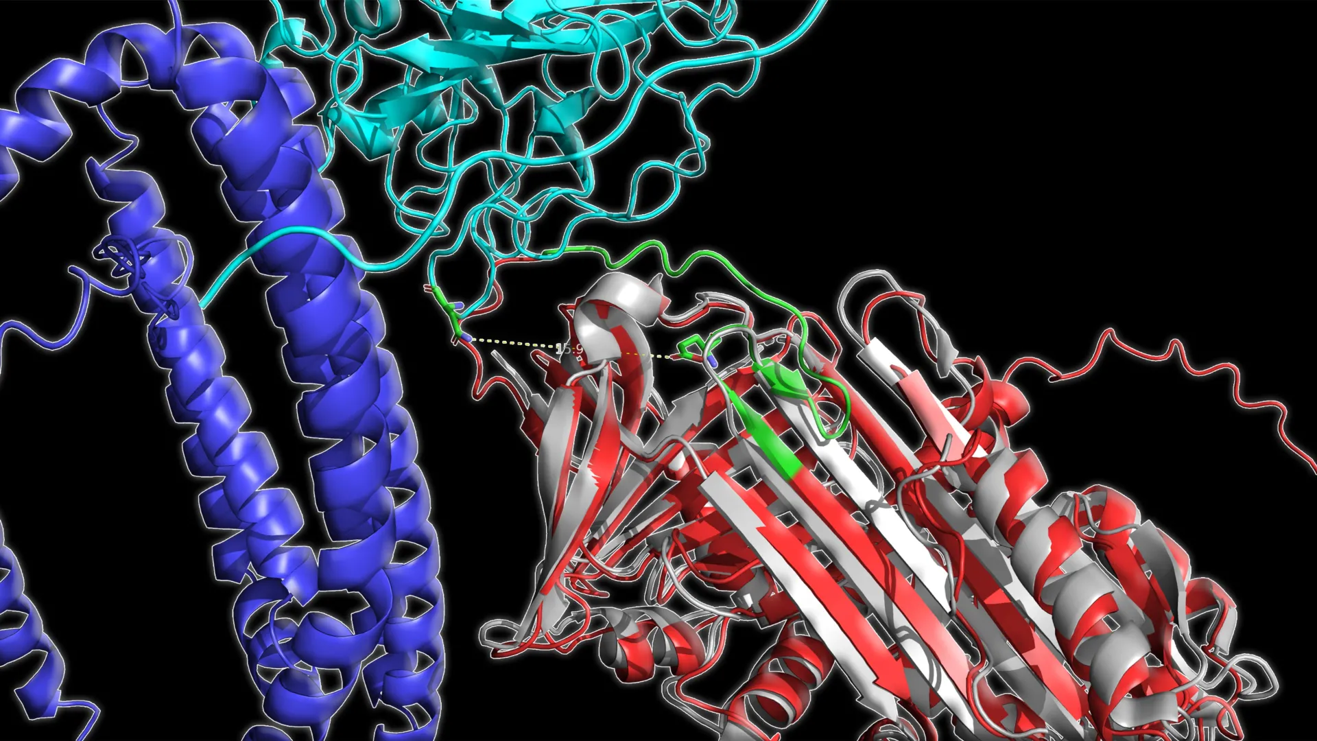
The researchers employed a highly sensitive analytical technique, mass spectrometry, to meticulously probe the structural landscape of the proteins present in the plasma samples. This method allowed them to discern the accessibility of specific amino acid residues within protein molecules, effectively revealing changes in their three-dimensional folding. Following the structural characterization, the team leveraged the power of machine learning algorithms. These advanced computational tools were instrumental in identifying subtle yet significant patterns within the protein structural data that were demonstrably associated with the different disease states.
The analytical findings were striking, revealing a consistent and discernible pattern across all participant groups. As Alzheimer’s disease progressed, a distinct shift was observed: certain proteins within the plasma exhibited a reduction in their structural "openness," indicating a more compact or altered configuration. Crucially, these observed structural alterations proved to be more discriminative for identifying the stage of the disease than traditional methods that solely focus on measuring the absolute concentration of specific proteins. This underscored the diagnostic potential of probing protein conformation rather than just abundance.
Identification of Three Key Proteins Correlated with Alzheimer’s Progression
Among the extensive array of proteins analyzed in the plasma samples, three specific proteins emerged with the most pronounced and significant associations with Alzheimer’s disease status. These proteins included C1QA, a component of the immune system known to play a role in immune signaling pathways; clusterin, a chaperone protein implicated in protein folding processes and the clearance of amyloid aggregates; and apolipoprotein B, a critical lipoprotein responsible for transporting fats in the bloodstream and contributing to the maintenance of vascular health. The involvement of these diverse proteins highlights the multifaceted nature of Alzheimer’s pathology.
Casimir Bamberger, a senior scientist at Scripps Research and a co-author of the study, expressed his astonishment at the findings, remarking, "The correlation was amazing." He further elaborated on the unexpected nature of their discovery: "It was very surprising to find three lysine sites on three different proteins that correlate so highly with disease state." The identification of specific "lysine sites" – particular locations within these proteins – that exhibited these strong correlations offered precise molecular targets for their diagnostic approach.
The ability of these identified structural changes at specific sites within these three proteins to accurately classify participants into one of the three diagnostic categories (cognitively normal, MCI, or Alzheimer’s) was remarkable, achieving an overall accuracy of approximately 83%. When the analysis was narrowed down to comparing just two groups at a time, such as distinguishing between healthy individuals and those with MCI, the accuracy rates soared above an impressive 93%. This level of precision suggests a powerful new avenue for diagnostic refinement.
Longitudinal Validation and Clinical Relevance of the Protein Structure Model
The robustness of the three-protein structural model was further validated through its application to independent cohorts of participants, reinforcing the generalizability of the findings. Moreover, the model demonstrated its reliability when researchers analyzed blood samples that had been collected from the same individuals at different points in time, spanning several months. This longitudinal analysis was critical for assessing the model’s ability to track disease progression.
In these repeat testing scenarios, conducted months apart, the protein structure panel consistently identified the disease status with an accuracy of approximately 86%. Critically, the model also proved capable of reflecting changes in an individual’s diagnosed status over time, suggesting its potential utility in monitoring the dynamic nature of the disease. Beyond disease classification, the derived "structural score" also exhibited a strong correlation with performance on standard cognitive assessments and a more moderate, yet significant, association with measurements of brain atrophy obtained through Magnetic Resonance Imaging (MRI).
Taken collectively, these compelling findings strongly indicate that the analysis of protein structural configurations in blood holds substantial promise as a complementary diagnostic tool to existing amyloid and tau-based assays. By focusing on structural alterations that are intimately linked to the underlying biological mechanisms of Alzheimer’s disease, this novel method could significantly enhance the ability of researchers and clinicians to accurately identify different disease stages, meticulously monitor disease progression, and objectively evaluate the efficacy of emerging therapeutic interventions.
Future Directions and Therapeutic Imperatives
The critical importance of early detection in the fight against Alzheimer’s disease cannot be overstated. Professor Yates emphasizes this point, stating, "Detecting markers of Alzheimer’s early is absolutely critical to developing effective therapeutics." He further posits that "If treatment can start before significant damage has been done, it may be possible to better preserve long-term memory." This sentiment underscores the transformative potential of an early diagnostic tool for improving patient outcomes.
Before this promising blood test can be widely implemented in clinical settings, further rigorous investigation is imperative. Larger-scale studies, characterized by extended follow-up periods, are necessary to conclusively confirm these initial findings and solidify their clinical validity. Furthermore, the research team is actively exploring the broader applicability of their protein structural profiling methodology. They are investigating whether this innovative approach can be adapted to identify similar molecular signatures in other challenging diseases, including Parkinson’s disease and various forms of cancer, potentially unlocking new diagnostic and therapeutic avenues across a spectrum of human ailments.
The seminal study, titled "Structural signature of plasma proteins classifies the status of Alzheimer’s disease," was authored by a collaborative team including Ahrum Son, Hyunsoo Kim, and Jolene K. Diedrich from Scripps Research; Heather M. Wilkins, Jeffrey M. Burns, Jill K. Morris, and Russell H. Swerdlow from the University of Kansas Medical Center; and Robert A. Rissman from the University of California San Diego. Financial support for this groundbreaking research was generously provided by the National Institutes of Health through grants RF1AG061846-01, 5R01AG075862, P30AG072973, and P30-AG066530.
About the Author


