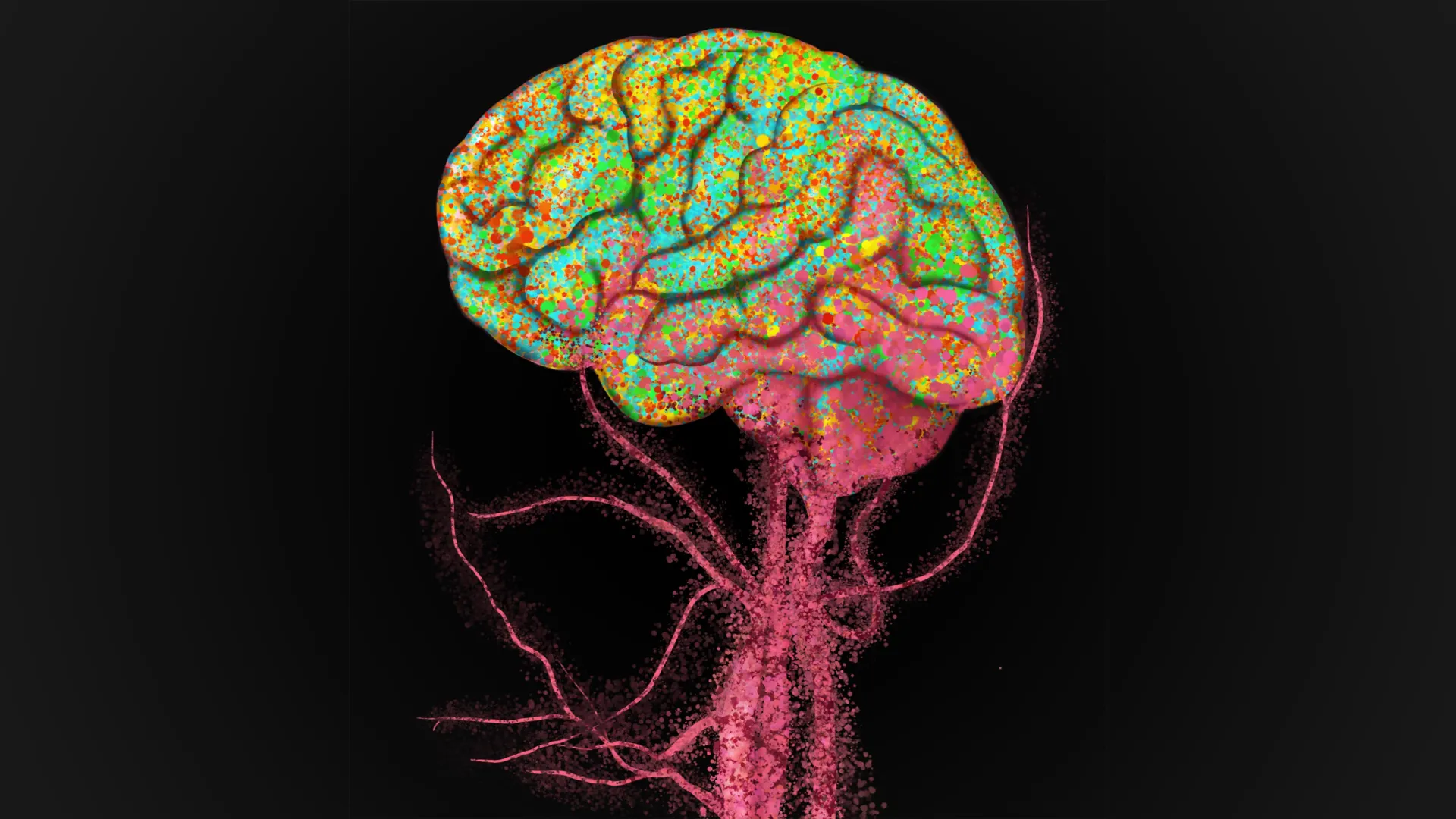
For a considerable period, the prevailing scientific consensus maintained a distinct separation between the brain’s intrinsic immune defenses and the immunological surveillance mechanisms operating throughout the rest of the body. This established view was largely underpinned by the existence of specialized immune cells within the central nervous system, coupled with the formidable physiological barrier known as the blood-brain barrier, which strictly curtails the passage of numerous substances and cellular entities into neural tissue. However, recent groundbreaking investigations originating from Stanford University are fundamentally challenging this long-held paradigm, proposing a more interconnected immunological landscape within the aging human brain.
The core finding of this research indicates that a substantial influx of immune cells, originating from the peripheral circulation, actively penetrates the human brain as individuals advance in age. This revelation has the potential to profoundly reshape the scientific community’s comprehension of the aging process within the brain and may pave the way for innovative therapeutic avenues for a spectrum of neurological disorders. The comprehensive study, which benefited from partial funding through the Knight Initiative for Brain Resilience at the Wu Tsai Neurosciences Institute, was recently disseminated in the esteemed scientific journal Nature.
Dr. Julia Belk, a postdoctoral scholar in pathology at Stanford Medicine and the lead author of the study, articulated the shift in perspective, stating, "We typically conceptualize the brain as an isolated entity. Our findings, however, reveal a significant migration of immune cells into the human brain during the aging process." This sentiment underscores a departure from the long-standing notion of the brain as a secluded immunological sanctuary.
The journey into this specific area of brain research was not a direct path for Dr. Belk. Her initial fascination with neuroscience emerged during her doctoral studies in the Department of Computer Science at Stanford’s School of Humanities and Sciences. During this period, she also underwent specialized training through Sarafan ChEM-H’s Chemistry/Biology Interface Predoctoral Training Program. She credits this formative experience with fostering a crucial interdisciplinary approach, seamlessly integrating fundamental scientific principles, computational methodologies, and clinical medicine.
This unique academic trajectory ultimately facilitated a pivotal collaboration with Dr. Siddhartha Jaiswal, a senior author of the current study, an associate professor of pathology at Stanford Medicine, and an integral member of the Institute for Stem Cell Biology and Regenerative Medicine. Their prior work had involved an extensive analysis of genetic data from thousands of individuals, some of whom had been under longitudinal observation for decades. In that preceding research, the team observed a notable correlation: individuals possessing specific clones of immune cells, generated by mutated blood stem cells, exhibited a significantly reduced likelihood of developing Alzheimer’s disease. This intriguing observation prompted the hypothesis that these unusual immune cells might be engaging with the brain in some capacity.
Subsequent investigations provided compelling evidence suggesting that some of these mutated cells could indeed infiltrate the brain itself. The mutations in question are linked to a condition known as clonal hematopoiesis of indeterminate potential, which is present in only a fraction of the population. Nevertheless, this discovery spurred the researchers to pose a broader, more encompassing question: Could the infiltration of immune cells from the bloodstream be a routine occurrence in aging humans?
Dr. Jaiswal elaborated on the traditional understanding, noting, "Unlike the majority of immune cells, which are continuously regenerated by blood stem cells originating from the bone marrow, it was generally presumed that immune cells within the brain self-renew throughout an individual’s lifespan, without any external contribution. Our initial findings suggest that this assumption may not always hold true."
This new research directly confronts a foundational concept regarding the brain’s resident immune cells, known as microglia. For many years, the prevailing scientific belief held that these specialized brain immune cells were established during embryonic development and subsequently constituted a self-sustaining population, requiring no replenishment from external sources throughout life. Under this established model, the migration of immune cells from the body’s systemic circulation into the brain and their integration into this resident population was not anticipated.
However, Dr. Belk and her colleagues began to entertain an alternative hypothesis. If peripheral immune cells could, in fact, enter the brain in certain individuals, it was plausible that this process might not be an anomaly but rather an integral and common feature of human aging. The notion that immune cells circulating in the blood could play a role in Alzheimer’s disease was both unconventional and a subject of considerable debate within the scientific community. In 2022, Dr. Jaiswal and his research group sought and received support from the Knight Initiative for Brain Resilience, an initiative dedicated to fostering novel approaches to studying brain resilience and neurodegenerative diseases.
With the assistance of an Innovation Award from the Knight Initiative, Dr. Belk, Dr. Jaiswal, and Dr. Howard Chang, a co-senior author and distinguished professor of genetics and cancer research at Stanford Medicine, embarked on an investigation into the reasons behind the observed increased resilience to Alzheimer’s in individuals with peripheral immune cells exhibiting specific characteristics. Before directly addressing that complex question, however, their immediate priority was to definitively ascertain whether immune cells originating from the bloodstream could indeed contribute to the microglia population within the brain.
To rigorously investigate this phenomenon, the researchers meticulously examined human brain tissue samples. They utilized specimens obtained from the Stanford Rapid Autopsy Center, a facility directed by co-author Dr. Jody Hooper, a professor of pathology at Stanford Medicine, and also incorporated samples from the University of Washington’s Alzheimer’s Disease Sequencing Project. These crucial programs collect both blood and post-mortem brain tissue from individuals who either had Alzheimer’s disease or were free from it. This unique dual-source approach provided the research team with an exceptional opportunity to conduct a direct comparison between the immune cells present in the bloodstream and those identified within brain tissue after death.
A significant methodological challenge lay in accurately determining the origin of the immune cells found within the brain. Because immune cells undergo continuous division, the scientists needed a reliable method to trace their cellular lineage, or "family trees." Their objective was to distinguish between cells that were descendants of the initial microglia population established at birth and those that had originated from blood stem cells in the bone marrow at later stages of life.
The researchers devised an ingenious solution by comparing the DNA of immune cells extracted from blood with the DNA of immune cells obtained from brain tissue. They employed shared genetic mutations as biological markers, analogous to how consumer ancestry testing services trace familial connections. Random mutations naturally accumulate in blood stem cells over the course of an individual’s life. Immune cells subsequently produced by these mutated stem cells inherit these identical mutations. Therefore, if two distinct populations of immune cells share the same mutations, it is highly probable that they share a common ancestral origin.
"If we observe the identical mutations in both the blood and the brain’s microglia, then we can be highly confident that the immune cells within the brain are indeed descendants of those found in the blood," Dr. Belk explained, highlighting the power of this genetic tracking method.
Leveraging this sophisticated approach, alongside advanced techniques developed during their prior 2023 research, Dr. Belk and her colleagues meticulously compared immune cells from paired blood and brain samples. The genetic signatures remarkably aligned, providing definitive evidence that immune cells from the peripheral circulation had successfully entered the brain. This infiltration process was observed to occur even in middle age, indicating it is not an exclusively late-stage phenomenon.
Further experimental analyses yielded another striking observation: once these peripheral immune cells successfully navigated into the brain, they underwent a remarkable transformation, differentiating into specialized microglia. The researchers also noted that this specific process of peripheral immune cell infiltration and subsequent differentiation into microglia does not appear to occur in other species, such as mice or non-human primates, suggesting it may be a uniquely human feature of brain aging.
Beyond fundamentally challenging established concepts of brain immunology, this groundbreaking discovery holds significant promise for the development of novel therapeutic strategies targeting the brain. "Now that we have confirmed that these immune cells can indeed enter the brain, we can explore a wide array of innovative engineering approaches to harness these peripheral immune cells for beneficial functions," Dr. Jaiswal remarked.
One compelling therapeutic possibility involves genetically engineering these circulating immune cells to specifically target and degrade the amyloid and tau protein aggregates, which are pathological hallmarks associated with neurodegenerative diseases like Alzheimer’s. Such engineered cells could potentially be administered prophylactically to individuals, intervening before the accumulation of these damaging protein deposits begins.
Moreover, this discovery has the potential to broaden the scope of research into the intricate relationship between the health and history of blood stem cells and their impact on the brain. Given that a substantial proportion of microglia in aging humans appear to originate from blood stem cells, any factor that influences the cells within the blood or bone marrow could consequently exert an influence on the brain’s immunological environment.
"Our findings strongly suggest that the life history of blood stem cells could significantly affect an individual’s susceptibility to brain diseases by altering the composition and function of microglia," Dr. Jaiswal concluded. For Dr. Belk, the implications of these results are particularly exciting, as they illuminate an aspect of brain aging that appears to be distinctively human. "I find this discovery to be incredibly exciting because it reveals a uniquely human characteristic of aging that we were previously unaware of," she stated.
About the Author