A groundbreaking scientific revelation from Weill Cornell Medicine and the Hospital for Special Surgery has pinpointed a previously uncharacterized population of stem cells, now understood to be the fundamental progenitors of the body’s tendons and ligaments – the vital connective tissues anchoring muscles to bones and providing structural integrity. This significant discovery not only sheds light on the intricate biological mechanisms governing musculoskeletal development but also offers a compelling new perspective on a prevalent and debilitating condition: lumbar spinal stenosis.
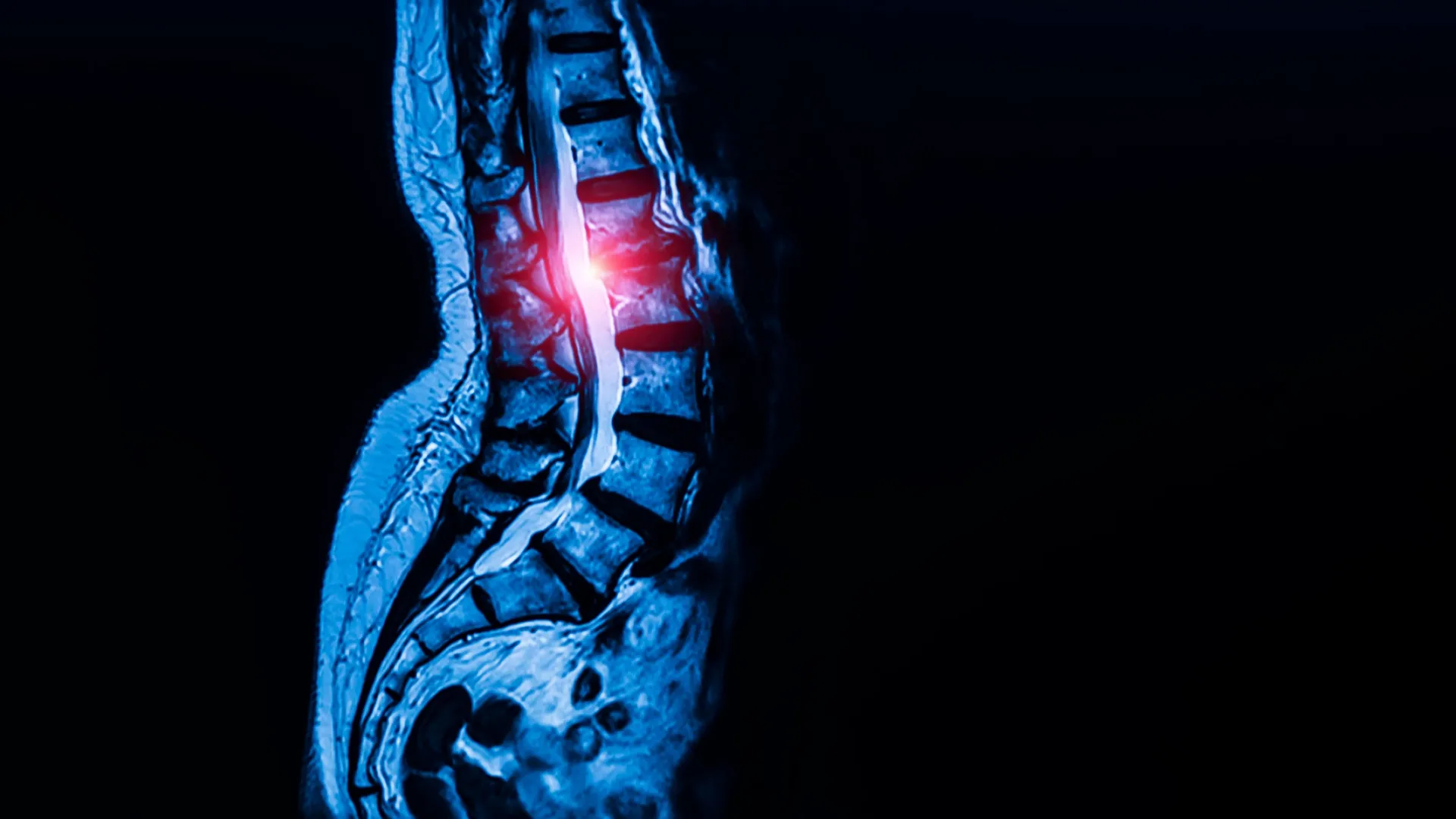
Lumbar spinal stenosis, a condition affecting an estimated 103 million individuals globally, is characterized by the progressive narrowing of the spinal canal, primarily due to the thickening of ligamentous structures within the lower back. This constriction exerts undue pressure on the delicate nerve roots housed within the canal, manifesting in a cascade of debilitating symptoms that can include persistent pain, sensory disturbances such as numbness or tingling, and profound difficulties with ambulation. The impact on an individual’s quality of life can be substantial, often leading to limitations in daily activities and a significant reduction in mobility.
The research, meticulously detailed in the esteemed scientific journal Cell on September 7th, proposes a paradigm shift in our understanding of spinal stenosis by identifying these foundational stem cells as potential instigators of the pathological processes underlying the condition. This opens up an exciting new frontier in therapeutic development, suggesting that interventions aimed at modulating the activity of these specific stem cells could pave the way for novel and more effective treatments for spinal stenosis, particularly for patients who currently face limited options beyond invasive surgical procedures once their condition reaches an advanced stage.
Dr. Matthew Greenblatt, a key investigator in this study and a distinguished figure in pathology and laboratory medicine at Weill Cornell, articulated the significance of this finding. He explained that while prior research had posited various candidate stem cell types responsible for connective tissue formation, none had definitively demonstrated the dual capacity for self-renewal and the generation of the complete array of mature cell types required for robust tendon and ligament development. "While previous studies had proposed several candidate stem cells, none had definitively shown that a single cell population could both self-renew and generate the full spectrum of tendon and ligament cell types," Dr. Greenblatt stated, emphasizing the unique and comprehensive role of the newly identified cells.
Following the identification of these specialized stem cells, the research team embarked on an investigation to determine their potential involvement in the pathogenesis of lumbar spinal stenosis. This exploration was particularly motivated by the limited therapeutic avenues available for patients whose stenosis has progressed to a severe degree, often necessitating surgical intervention to alleviate nerve compression.
Dr. Sravisht Iyer, an associate professor of orthopedics at Weill Cornell and a practicing spine surgeon at the Hospital for Special Surgery (HSS), highlighted the transformative potential of this discovery for patient care. "Identifying these specialized stem cells unlocks a new area of research that allows us to address this disease much more mechanistically, rather than just waiting until a patient’s condition worsens and requires surgery to relieve the nerve compression," Dr. Iyer remarked. He further elaborated on the profound implications for the future of spinal care, stating, "The findings are exciting for their potential to change the way we deliver spinal care."
The quest to isolate and characterize the stem cells responsible for constructing tendons and ligaments was a complex undertaking. Dr. Greenblatt and his team brought a wealth of prior experience in identifying crucial skeletal stem cell populations, having previously elucidated the cells involved in fracture repair within the bone’s outer layer and those contributing to the formation of the skull and spine. However, pinpointing an analogous cell for tendons and ligaments proved to be a considerably more formidable challenge.
The inherent difficulty stemmed from the cellular composition of these connective tissues. Unlike bone, tendons and ligaments are characterized by a high density of fibroblast-like cells, which exhibit a remarkable degree of morphological similarity. This uniformity made it exceptionally challenging to discern and isolate distinct cell populations based on visual inspection alone.
To overcome this obstacle, the researchers employed sophisticated single-cell analysis techniques. "We analyzed thousands of individual cells and sorted them into individual cell types. Then we identified which one had the properties we associate with ‘stemness,’" Dr. Greenblatt explained, detailing the meticulous process of cell sorting and characterization. The concept of "stemness" in this context refers to a critical attribute: the capacity of a rare cell population to perpetuate itself through self-replication while simultaneously differentiating into the specialized mature cells essential for the continuous maintenance and repair of tendon and ligament tissues.
In preclinical studies conducted in mice, these foundational stem cells were found to reside within specific niches within tendons and ligaments, suggesting these locations serve as vital reservoirs supporting tissue growth and regeneration. The insights gained from these animal models were then meticulously applied to search for analogous cells in human subjects. Crucially, human ligament samples utilized in the study were obtained from tissue resected during surgical procedures performed by Dr. Iyer, with the full informed consent of the patients.
Under the leadership of Dr. Lingling Hu, a postdoctoral fellow in both Dr. Greenblatt’s and Dr. Iyer’s laboratories, the research team successfully validated that the human cells possessed the requisite stem cell characteristics: the ability to self-renew and the capacity to generate mature ligament cells. Their investigations further revealed that these universal stem cells for tendons and ligaments were not confined to the spine but were distributed throughout the body.
"We looked in the kneecap ligament; we looked at the Achilles tendon; and everywhere we looked, we found this cell," Dr. Greenblatt stated, underscoring the widespread presence and fundamental role of these cells. "So, we think this is the universal stem cell for tendons and ligaments throughout the body." This broad distribution highlights their pervasive importance in maintaining the integrity of the musculoskeletal system.
The subsequent phase of the research focused on investigating the potential link between these newly identified stem cells and the development of lumbar spinal stenosis. The scientists compared stem cells harvested from individuals diagnosed with spinal stenosis to those collected from spinal ligaments of patients who had undergone surgery for herniated discs but did not exhibit any symptoms of stenosis. Rigorous informed consent protocols were adhered to for all patient samples.
A key finding emerged: ligaments from individuals suffering from spinal stenosis harbored a significantly higher abundance of these specific stem cells compared to their healthy counterparts. Furthermore, when these stem cells were transplanted into a mouse model, they demonstrated a pronounced propensity to generate tendon cells, a characteristic more pronounced than in stem cells derived from individuals without stenosis.
"Though spinal stenosis is a complex condition, this really showed us that these cells are contributing to the pathology," Dr. Greenblatt affirmed, acknowledging the multifaceted nature of the disease while emphasizing the direct contribution of these stem cells. His affiliation with the Sandra and Edward Meyer Cancer Center at Weill Cornell adds another layer of expertise to his research, particularly concerning cellular proliferation and control mechanisms.
Delving deeper into the intracellular mechanisms, the researchers observed a notable difference in calcium signaling pathways within the stem cells associated with stenosis. These cells exhibited heightened calcium signaling activity when contrasted with healthy stem cells. Calcium signaling plays a pivotal role in intracellular communication, orchestrating critical cellular processes such as growth and metabolic activity.
Intriguingly, the researchers were able to experimentally induce excessive tissue growth in stem cells from healthy ligaments by genetically enhancing their calcium signaling. This demonstration provided compelling evidence for the direct role of aberrant calcium signaling in driving pathological tissue proliferation.
Conversely, the study revealed that inhibiting calcium signaling in a mouse model of lumbar spinal stenosis effectively curbed abnormal cell growth. This observation strongly suggests that calcium signaling pathways represent a promising therapeutic target for the management of spinal stenosis. The findings also raise the tantalizing prospect that calcium channel blockers, a class of pharmaceuticals already widely prescribed for the treatment of hypertension, could potentially be repurposed or investigated as a therapeutic strategy for spinal stenosis. However, the researchers stressed that extensive clinical trials will be indispensable to ascertain the safety and efficacy of such an approach in human patients.
"This is probably the first work that’s shown a potential therapeutic target for one of the most common spine conditions in the world," Dr. Iyer stated, underscoring the novelty and clinical relevance of their findings.
The implications of this research extend far beyond the immediate scope of spinal stenosis. Dr. Greenblatt indicated plans to explore whether these same stem cells are implicated in other connective tissue disorders, such as Marfan syndrome, a genetic condition that affects the body’s connective tissues across multiple organ systems.
Moreover, this discovery holds the potential to significantly advance our understanding of why certain tendon and ligament injuries prove notoriously difficult to heal. Future research avenues may encompass a deeper investigation into conditions like rotator cuff tears, Achilles tendon injuries, the intricacies of ligament reconstruction, and the underlying mechanisms of chronic tendon degeneration.
"Given that this cell appears to be the ultimate origin of all tendon and ligament cells, defects in this cell are likely at the heart of a wide range of tendon and ligament disorders," Dr. Greenblatt concluded, highlighting the fundamental and far-reaching impact of their foundational discovery on the broader field of connective tissue biology and pathology.
This significant research endeavor was made possible through substantial support from various esteemed organizations, including the Marfan Foundation’s Victor A. McKusick Fellowship, a Kellen Scholars Award, the Children’s Tumor Foundation, the Arthritis National Research Foundation, a Jumpstart award from Weill Cornell, a National Research Foundation of Korea award, multiple National Institutes of Health grants, a MIND Prize from the Pershing Square Foundation, a Mary Kay Ash Foundation Award, an Innovator Award from the Marfan Foundation, and a Burroughs Wellcome Career Award for Medical Scientists.
About the Author