New molecular map reveals how the flu virus hijacks human cells
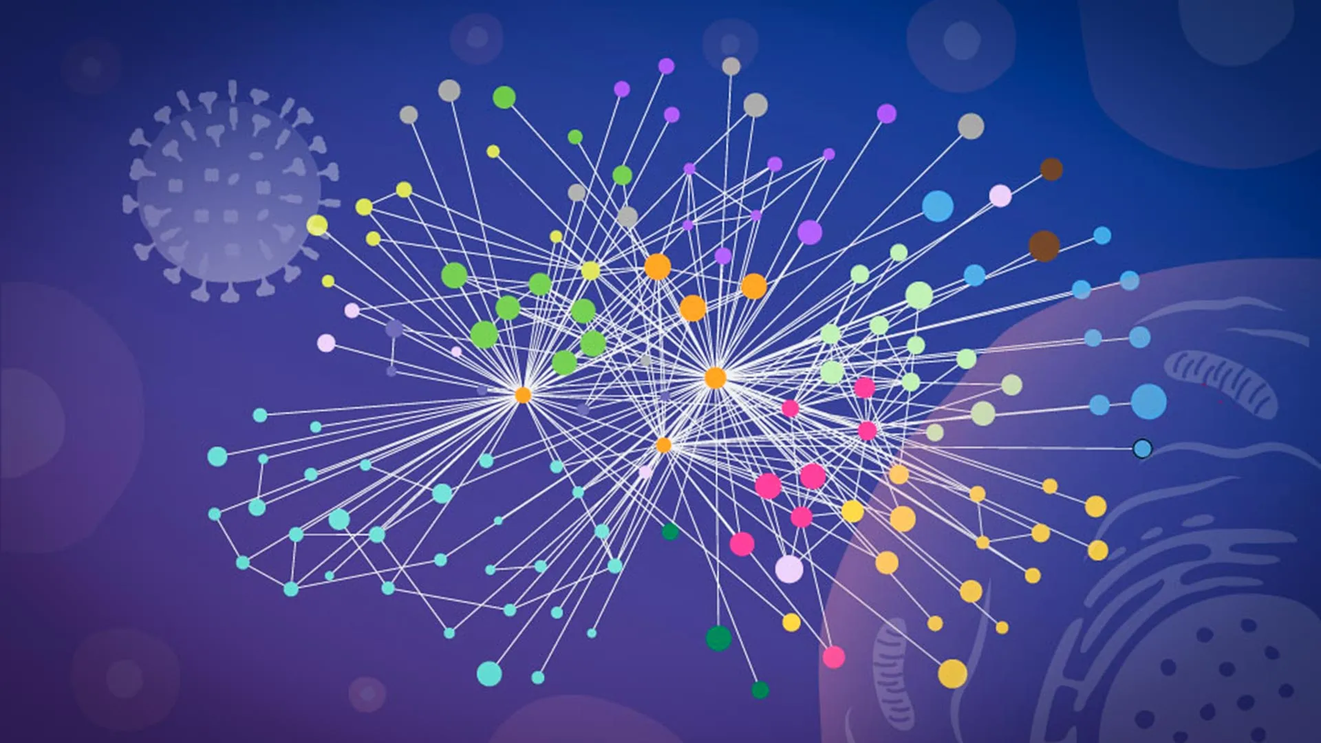
The pervasive impact of seasonal influenza cannot be overstated, with millions of individuals worldwide succumbing to severe illness annually, and the virus being implicated in hundreds of thousands of fatalities. Influenza A, in particular, has a notorious history, having been the progenitor of devastating pandemics, including the calamitous Spanish Flu outbreak of 1918. The insidious nature of the virus lies in its ability to infiltrate a host cell and, upon entry, liberate its genetic material – RNA – which serves as the blueprint for synthesizing a select group of viral proteins. These proteins then disseminate throughout the cellular interior, effectively commandeering the host cell’s sophisticated molecular machinery and transforming it into a factory dedicated to the mass production of new viral progeny.
A comprehensive understanding of this cellular invasion process is paramount for the development of more potent influenza vaccines and the creation of novel antiviral therapeutics. To achieve this crucial objective, scientists must precisely identify which viral proteins engage with human proteins, pinpoint the exact locations where these critical encounters transpire, and elucidate the specific mechanisms by which the virus exploits these interactions to facilitate its own replication. The present study represents a significant leap forward, being the first large-scale mapping of direct physical contacts between influenza viral proteins and their human cellular counterparts within undisturbed, infected cells. The remarkable precision of the structural detail achieved allows for the modeling of how these interacting proteins are likely to interlock.
Jan Kosinski, a distinguished Group Leader at EMBL Hamburg and the Centre for Structural Systems Biology (CSSB), highlighted the transformative potential of their work, stating, "Our research establishes a novel paradigm for investigating flu-host interactions within their native context and with profound structural insight." He further elaborated that the current findings provide a static snapshot of a critical juncture during the infection cycle, thereby opening avenues for longitudinal studies of flu-host interactions across the entire duration of the infection.
The challenge of precisely tracking protein-protein interactions during an active viral infection is notoriously formidable. Historically, numerous studies have relied on biochemical techniques that necessitate the disruption of cellular integrity – essentially breaking open the cells – before any measurements of protein contacts could be made. This destructive process, however, carries a significant drawback: it can distort the authentic cellular landscape. Once the intricate internal compartments of a cell are dismantled, proteins that were previously segregated may come into proximity in the laboratory setting, leading to artificial interactions. Conversely, transient, weak, or location-specific interactions that are vital to the infection process can be lost. Consequently, researchers have often faced difficulties in discerning which molecular connections were genuinely operative during the natural course of infection.
"It was at this juncture that we discovered our collaborators, Boris Bogdanow and Fan Liu, at the FMP Berlin, had ingeniously adapted cross-linking mass spectrometry (XL-MS), a well-established technique for mapping protein contacts, into a specialized version specifically engineered for virus-infected cells," explained Kosinski. This bespoke methodology provided the critical breakthrough the research team had been seeking. It conferred the ability to capture interactions that are fleeting in duration or confined to particular subcellular compartments within an infected cell.
Boris Bogdanow, now a Junior Research Group Leader at the Institute of Virology at Charité — Universitätsmedizin Berlin, underscored the significance of their refined approach, stating, "XL-MS enables us to capture protein-protein interactions directly within infected, intact cells, while simultaneously furnishing structural information regarding the nature of these interactions." He further elaborated that this capability offers invaluable insights into the molecular interfaces between the virus and the human cell, with the potential, through structural modeling, to identify actionable targets for future pharmaceutical interventions.
The researchers ingeniously integrated their experimentally derived XL-MS data with advanced computational structural modeling techniques. This synergistic approach enabled them to not only identify the specific viral and human proteins that engage in direct interaction but also to estimate their precise spatial orientation at the moment of contact. For the construction of these intricate structural models, the team leveraged a modified iteration of AlphaFold, the celebrated protein structure prediction algorithm that has revolutionized the field of structural biology.
"The paramount advantage of our modified AlphaFold approach lies in its capacity to directly incorporate our experimental cross-linking data into the structural modeling process," elucidated Kosinski. "This crucial step guides the model by indicating which segments of viral and host proteins are in close proximity within infected cells. This integration proved particularly invaluable for deciphering virus-host complexes, which are notoriously challenging to predict with high accuracy using conventional methods."
The comprehensive findings, published in the prestigious journal Nature Microbiology, have illuminated two particularly noteworthy strategies that influenza A employs in its systematic takeover of host cells. The first strategy centers on hemagglutinin, a key protein embedded in the virus’s outer membrane. Influenza utilizes hemagglutinin as its primary vehicle for attaching to and subsequently invading host cells. The research team meticulously tracked the journey of this protein as it navigated the cell’s complex internal transport and processing network. This network, a sophisticated system of membrane-bound compartments, is responsible for folding, modifying, and preparing proteins before their directed transport to their ultimate cellular destinations. The detailed analysis revealed that a number of host cell proteins play a crucial role in the proper folding and modification of hemagglutinin during the infection process. Intriguingly, some of these host proteins were previously known to possess poorly understood functions, suggesting a novel role for them in the context of viral infection.
The second pivotal discovery pertained to paraspeckles, which are small, droplet-like structures found within the cell’s nucleus. The study unequivocally demonstrated that infection with influenza A triggers the dissolution of these paraspeckles. Upon their disintegration, paraspeckles release RNA-binding proteins that were previously sequestered within them. The virus can then potentially harness these liberated proteins to bolster its own replication efforts.
Iuliia Kotova, a former predoctoral fellow in the Kosinski Group at EMBL Hamburg and the first author of the publication, expressed her surprise at this particular finding: "What astonished us most were the paraspeckles. Witnessing these minuscule nuclear organelles consistently dissolve across every cell line and every influenza strain we examined strongly suggested that this is not merely a byproduct of infection, but rather a deliberate strategy." This disruption likely confers multiple advantages upon the virus.
Kosinski further hypothesized about the potential secondary benefits for the virus, noting, "There may also be a secondary advantage for the virus: some evidence indicates that paraspeckles contribute to cellular stress responses and the regulation of antiviral genes. Therefore, their disruption could simultaneously compromise components of the cell’s defense mechanisms."
The successful execution of this ambitious project was made possible through the synergistic collaboration and shared resources of three distinct institutions. The critical cross-linking mass spectrometry analyses were conducted at Charité in Berlin, while sophisticated glycoproteomics analyses were performed at the EMBL Proteomics Core Facility. The complex AlphaFold modeling was carried out on the robust EMBL Compute Cluster, and the essential microscopy imaging was undertaken at the Advanced Light and Fluorescence Microscopy (ALFM) Facility at CSSB.
This groundbreaking research fundamentally alters our perception of how to study viral pathogens, particularly those with pandemic potential. The findings underscore the immense value of examining molecular contacts within the intact infected cell to comprehensively understand both the location and the precise mechanisms by which a virus commandeers human cellular machinery. The researchers posit that this ‘mapping in context’ approach holds significant promise for elucidating the operational strategies of a wide range of other viruses.
Kosinski emphasized the broad applicability of their methodology, stating, "While the specific host factors and mechanisms often vary from one virus to another, we believe our overall approach – a combination of in-cell cross-linking, structural modeling, and targeted cell-biology follow-up to map native virus-host interactions at specific stages of infection – remains broadly applicable." Although the current study utilized a laboratory-adapted strain of influenza, the researchers are confident that their innovative strategy can be extended to investigate viruses with a higher propensity for causing pandemics. Bogdanow concurred, adding, "Although this study has focused on a lab-adapted strain, it lays the essential groundwork for applying this methodology to viruses of potential pandemic relevance, such as H5N1, and for uncovering the intricate interaction networks that underpin their proliferation within human cells."

Related News