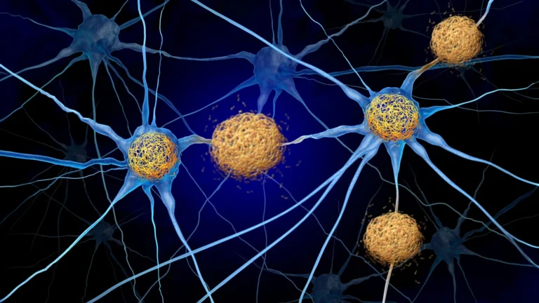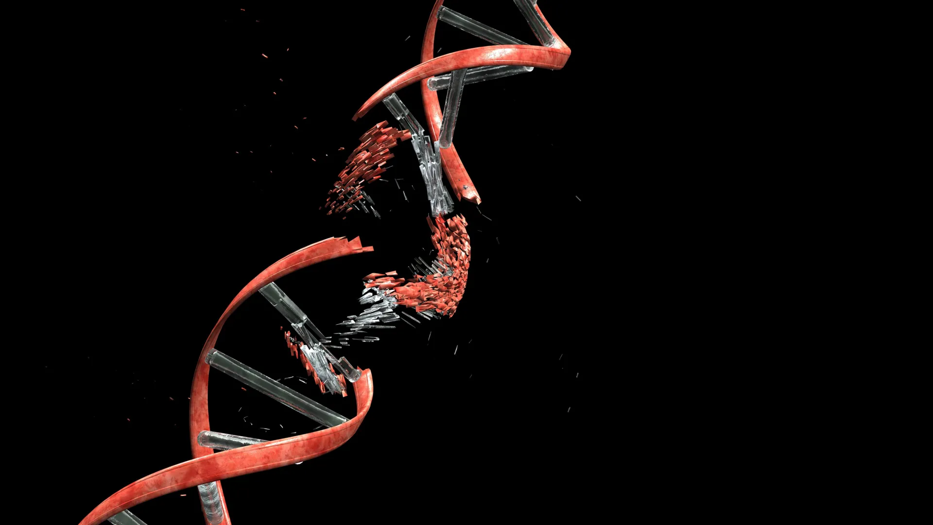
The intricate process of brain formation, particularly the journey of nascent nerve cells to their designated functional sites, involves a surprising and fundamental requirement: controlled fracturing of their own genetic material. This newly discovered mechanism, detailed in a groundbreaking study published in the esteemed journal Nature, challenges conventional understandings of cellular integrity and highlights a unique adaptive strategy employed by the developing central nervous system. Researchers from Kyoto University’s Institute for Integrated Cell-Material Sciences (WPI-iCeMS), in collaboration with several other leading academic institutions, have illuminated how the physical stresses encountered by migrating neurons during their critical developmental phase induce significant and, crucially, repairable DNA damage, specifically double-strand breaks.
These double-strand breaks, typically considered highly detrimental and harbingers of mutations, cellular malfunction, and even programmed cell death, have been identified not as accidental consequences but as an integral and orchestrated component of healthy cerebral cortex development. The study’s lead author, Professor Mineko Kengaku of WPI-iCeMS, emphasized that the developing brain possesses an exceptional capacity to both tolerate and efficiently mend this induced genetic damage. "Our findings suggest a profound evolutionary adaptation where the developing brain has engineered a mechanism to leverage a degree of controlled genomic instability for its own architectural needs," Professor Kengaku stated, underscoring the significance of understanding the boundaries of this tolerance and the implications of incomplete repair.
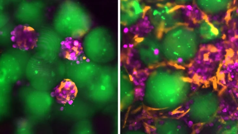
To meticulously dissect the origins and consequences of this phenomenon, the research team devised ingenious experimental models. They successfully replicated the formidable physical obstacles that migrating neurons confront by guiding these cells through precisely engineered microfluidic channels. These channels were designed to meticulously mimic the constrained and densely packed environments characteristic of a growing neural network. Employing advanced fluorescent labeling techniques, the scientists were able to visually track the emergence of double-strand DNA breaks in real-time as the neurons navigated these confined pathways. Remarkably, upon exiting the restrictive channels and reaching their simulated destinations, the observed DNA damage began to recede. The vast majority of these breaks were effectively repaired within a 24-hour period, allowing the neurons to resume normal physiological functions without apparent lasting repercussions.
The investigation pinpointed a key cellular player in this process: Topoisomerase IIα, an enzyme normally tasked with managing torsional stress within the DNA helix. Under typical cellular conditions, this enzyme acts like a skilled artisan, temporarily severing DNA strands to alleviate the tension generated by the dynamic activities of the cell, and then meticulously rejoining them. This can be conceptually understood as momentarily cutting a twisted electrical cord to untangle it before reconnecting the ends. However, the study revealed that when neurons are subjected to the intense mechanical pressures associated with squeezing through exceedingly narrow cellular interstices, Topoisomerase IIα can become transiently ensnared in the process, leaving behind severed DNA strands. In such instances, the neuron’s cellular machinery relies on a robust repair pathway known as non-homologous end joining to re-establish the continuity of the broken DNA fragments.
A crucial distinction emerged when comparing the DNA damage response of developing neurons to that of certain cancerous cells. When subjected to similar microchannel confinement, cancer cells exhibited a more indiscriminate pattern of DNA damage, frequently leading to disruptions in cellular operations or initiating apoptotic cascades. In stark contrast, the DNA breaks observed in developing neurons were notably localized to genomic regions that are not actively transcribed for essential protein synthesis. This strategic sparing of critical genes ensures that, despite the temporary genetic disruption, the neurons can maintain their fundamental functional integrity. This selective vulnerability and resilience underscore the sophisticated regulatory mechanisms at play during neurogenesis.
To further probe the ramifications of compromised DNA repair, the research team employed genetic engineering in a mammalian model. They created mice with a deficiency in Ligase 4, an enzyme indispensable for the non-homologous end joining repair pathway, specifically within their newly formed cerebellar neurons. Curiously, these mice exhibited normal development in their initial stages, showing no overt abnormalities. However, as they matured into adulthood, subtle yet progressive difficulties with balance control began to manifest. These observed motor deficits bear a striking resemblance to symptoms associated with certain human genetic disorders characterized by genomic instability, particularly those impacting the cerebellum, a brain region vital for motor coordination and balance.
These findings collectively suggest that the dynamics of DNA breakage and repair are far more pervasive and functionally significant in brain biology than previously appreciated. The scientific community is now poised to investigate whether these early-stage genomic alterations contribute to the subtle yet distinct variations observed among individual neurons and, more broadly, whether they play a role in the etiology of neurodevelopmental disorders and neurodegenerative conditions that emerge later in life. Professor Kengaku articulated this paradigm shift, stating, "This research fundamentally alters our perspective on the neuronal genome. While all neurons originate from a common genetic blueprint, the cumulative effects of DNA damage and subsequent repair mechanisms can introduce subtle genetic divergences between individual cells, imprinted by their unique mechanical journeys. It appears that elements of this developmental history are literally inscribed within the genome itself, shaping the future of neural circuitry and potentially influencing susceptibility to disease." The collaborative effort that produced these insights involved researchers from the University of Tokyo, Osaka University, the National University of Singapore, and the Tokyo Metropolitan Institute of Medical Science, alongside the lead institution, Kyoto University.
About the Author