Chronic liver disease represents a formidable global health challenge, leading to severe morbidity and mortality for millions. In the United States alone, over 10,000 individuals are currently on waiting lists for a liver transplant, a critical procedure that often serves as the last resort for end-stage hepatic failure. This urgent demand, however, is consistently thwarted by a severe scarcity of suitable donor organs. Furthermore, a significant subset of patients afflicted with severe liver dysfunction are medically ineligible for surgical transplantation due to their fragile health, rendering them too vulnerable to endure the rigors of such a complex operation. These profound limitations in conventional treatment avenues underscore the imperative for innovative therapeutic strategies.
Addressing this critical unmet need, a team of bioengineers from the Massachusetts Institute of Technology (MIT) has unveiled a groundbreaking approach: the development of injectable, cellularly-engineered constructs designed to furnish essential hepatic functionalities within the body. Termed "satellite livers" by their creators, these sophisticated cellular grafts aim to provide supplementary metabolic support, potentially revolutionizing how liver disease is managed. The initial findings, published in Cell Biomaterials, detail promising results from murine studies where the transplanted liver cells demonstrated sustained viability and metabolic activity for at least two months post-injection, consistently producing the enzymes and proteins characteristic of a healthy liver.
The research, led by senior author Professor Sangeeta Bhatia, the John and Dorothy Wilson Professor of Health Sciences and Technology and of Electrical Engineering and Computer Science at MIT, and a distinguished member of the Koch Institute for Integrative Cancer Research and the Institute for Medical Engineering and Science (IMES), marks a significant stride in regenerative medicine. Vardhman Kumar, an MIT postdoctoral researcher, served as the lead author on the pivotal study. Professor Bhatia articulates the vision behind this innovation: "We envision these as auxiliary hepatic modules. The ability to introduce these cells into the biological system while leaving the compromised native organ in situ offers a pathway to augment vital functions."
The liver, a quintessential organ, orchestrates a myriad of critical physiological processes, estimated to encompass nearly 500 indispensable biological roles. These include, but are not limited to, the intricate regulation of blood coagulation, the meticulous detoxification of the circulatory system from pathogens and toxins, and the complex metabolism of pharmaceutical compounds and nutrients. The bulk of these crucial tasks are performed by specialized parenchymal cells known as hepatocytes. For over a decade, Professor Bhatia’s laboratory has been at the forefront of investigating novel methodologies to reinstate hepatocyte activity without necessitating invasive surgical interventions. Previous strategies involved encapsulating hepatocytes within biocompatible materials, such as hydrogels, yet these still typically required surgical implantation. The pursuit of an injectable solution represents a leap forward, promising to circumvent the inherent challenges and risks associated with surgical procedures.
The core innovation of the current study lies in creating a highly optimized, engineered microenvironment that not only promotes the survival of transplanted cells but also facilitates the non-invasive assessment of graft viability and integration. The MIT team formulated an injectable suspension comprising hepatocytes and minute hydrogel microspheres. These micro-scale particulate biomaterial gels serve a dual purpose: they facilitate cellular cohesion, preventing dispersion of the transplanted cells, and critically, they encourage rapid integration with the host’s vasculature.
A key material property of these microspheres is their rheological behavior. When sufficiently concentrated and subjected to shear forces, the material exhibits fluid-like characteristics, enabling its passage through a standard syringe. Once delivered into the body, the material undergoes a phase transition, reverting to a stable, semi-solid gel state. This property is vital for creating a stable scaffold. Hydrogel microspheres have previously been explored in contexts such as wound healing, where their porous structure allows for cellular migration and the generation of new tissue. The MIT researchers ingeniously adapted this established technology to construct a stable, localized tissue graft capable of sustained function post-injection.
"Our approach leveraged this existing technology to forge a precisely tailored microhabitat for cellular engraftment," explains Kumar. He elaborates on the necessity of this engineered niche: "Without these spheres, direct injection of hepatocytes would likely result in inefficient host integration. These microspheres furnish the hepatocytes with a protective environment where they can remain localized and establish connections with the host’s circulatory system with significantly accelerated kinetics." The injectable matrix is further enriched with fibroblast cells, which are known for their supportive role in enhancing hepatocyte longevity and actively promoting neovascularization, the critical process of new blood vessel formation within the newly formed tissue.
To refine the delivery mechanism, the research team collaborated with Nicole Henning, an ultrasound research specialist at the Koch Institute. Together, they pioneered a technique for precisely delivering the cellular mixture using an ultrasound-guided injection system. This sonographic guidance ensures accurate placement of the graft. Moreover, ultrasound can subsequently be employed as a non-invasive diagnostic tool to longitudinally monitor the implant, assessing its stability, size, and overall health over time without requiring additional invasive procedures. This capability is paramount for clinical translation, offering a safe and repeatable method for tracking therapeutic efficacy.
The versatility of these cellular grafts extends to their potential placement within the body. In the murine experiments, the researchers strategically implanted the "mini livers" into the perigonadal adipose tissue, a fatty depot within the abdomen. Future iterations of this therapeutic intervention could potentially target other anatomical sites, including the splenic parenchyma or regions adjacent to the kidneys. Critically, the transplanted hepatocytes do not necessitate proximity to the patient’s native liver. Provided the graft is situated in an area with adequate spatial volume and a robust vascular supply, the engrafted hepatocytes demonstrate the capacity to replicate the metabolic activities of native hepatic cells, irrespective of their specific location. Kumar emphasizes this point: "For the vast majority of hepatic disorders, the therapeutic graft does not require anatomical proximity to the existing liver."
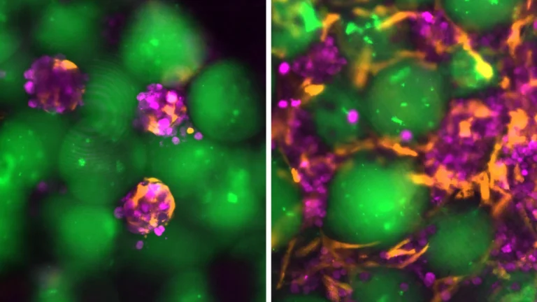
Upon delivery into the perigonadal adipose tissue during the mouse trials, the injected liver cells and microspheres coalesced into a robust, integrated tissue mass. Subsequently, progressive neovascularization permeated the implant, leading to the formation of new blood vessels that directly integrated with the graft. This critical vascularization established a localized trophic support system, supplying essential nutrients and oxygen, which proved instrumental in sustaining the viability and metabolic activity of the engrafted hepatocytes. "The nascent blood vessels formed in intimate association with the hepatocytes, which was fundamental to their survival," Kumar states. "This direct vascular access ensured efficient nutrient delivery, enabling them to function optimally and produce the specific proteins we anticipated." The liver cells maintained their functional integrity throughout the entire eight-week study period, continuously releasing specialized proteins into the animals’ circulation. This remarkable durability exhibited by the grafts suggests that the technique holds substantial promise for development into a protracted therapeutic application for chronic liver disease.
The researchers envision several transformative applications for this innovative technology. For certain patient populations, these injectable hepatic grafts could offer a viable alternative to complex surgical interventions. For others, they could serve as an interim therapeutic strategy, providing essential liver support while the patient awaits the availability of a suitable donor organ. "We conceptualize this technology as not only an alternative to surgical procedures but also as a crucial bridge to transplantation," Kumar elaborates. "These grafts can furnish vital support until a donor organ materializes. Moreover, should further therapy or additional grafts be required, the logistical and clinical hurdles associated with this injectable technology are substantially reduced compared to undergoing repeat surgeries."
A key consideration for the clinical implementation of this approach involves managing the host immune response. Currently, patients receiving such cellular transplants would likely require immunomodulatory pharmacotherapy to prevent their immune systems from rejecting the foreign cells. The team is actively investigating advanced strategies to circumvent this challenge, including the development of "immunogenically camouflaged" hepatocytes designed to evade immune detection. Another promising avenue involves leveraging the hydrogel microspheres themselves as localized delivery vehicles for immunosuppressive drugs, releasing these agents directly within the graft microenvironment to mitigate systemic side effects.
This pioneering research, supported by significant funding from the Koch Institute Support (core) grant from the National Cancer Institute, the National Institutes of Health, the Wellcome Leap HOPE Program, a National Science Foundation Graduate Research Fellowship, and the Howard Hughes Medical Institute, represents a beacon of hope for countless individuals grappling with liver failure. By offering a less invasive, potentially more accessible, and highly adaptable therapeutic option, these bioengineered cellular grafts could profoundly reshape the landscape of liver disease management, moving towards a future where organ scarcity is no longer an insurmountable barrier to life-saving treatment.
About the Author