Unraveling the Blueprint: Cellular Decision-Making Orchestrates Human Brain Architecture.
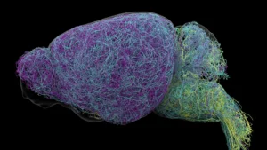
The intricate construction of the human brain, a process commencing long before birth, is governed by a sophisticated symphony of cellular choices. At the heart of this developmental marvel reside radial glial cells, a specialized category of progenitor cells instrumental in generating the myriad components that confer upon the human brain its distinctive complexity and size. These remarkable cells are the principal architects, responsible for producing the vast quantities of neurons and supportive glial cells that constitute the cerebral cortex, the brain’s command center for higher cognitive functions such as abstract thought, memory retention, and linguistic processing. Furthermore, radial glia are theorized to play a pivotal role in the disproportionately expansive growth of the human cortex when juxtaposed with that of other mammalian species. While the majority of these cells undergo programmed cell death prior to birth, a fascinating biological parallel emerges in the context of certain brain cancers, where similar cells can re-emerge, a phenomenon that continues to elude complete scientific comprehension.
Dr. Aparna Bhaduri, an assistant professor of biological chemistry at the David Geffen School of Medicine at UCLA, eloquently describes radial glia as "the coolest cells that have ever existed," emphasizing their critical contribution to what defines human cognition and behavior. She further elaborates that these cells are not only central to human uniqueness but are also implicated in the genesis of a spectrum of neurodevelopmental and neuropsychiatric disorders, as well as in oncological processes. Consequently, deciphering the mechanisms by which these cells make their developmental determinations is posited as a foundational step toward understanding the origins of these complex conditions.
Recent groundbreaking research, disseminated across two prominent scientific journals, Cell and Science, offers unprecedented insights into the intricate decision-making processes employed by radial glia during development. These studies, spearheaded by Dr. Bhaduri and her collaborators, reveal that these foundational cells integrate information from two fundamentally distinct sources: their internal metabolic state, reflecting how they process nutrients, and direct physical cues emanating from other developing brain regions. The convergence of these findings illuminates novel pathways by which the human cortex achieves its extraordinary diversity of neuronal and glial subtypes.
One of the pivotal discoveries, detailed in the Cell publication, involves the creation of a comprehensive metabolic atlas of the developing human cortex. This ambitious project, a collaborative effort between Dr. Bhaduri’s laboratory and that of Dr. Heather Christofk, was primarily driven by the meticulous work of co-first authors Jessenya Mil and Jose Soto. To construct this detailed map, the research team meticulously analyzed both donated human brain tissue and sophisticated brain organoids meticulously cultured from pluripotent stem cells. The findings from this extensive analysis led to a surprising and paradigm-shifting conclusion: metabolism is not merely a passive supporting element of brain development; rather, it actively exerts influence over the types of cells that are ultimately generated.
The researchers observed a pronounced dependence of radial glia on the pentose phosphate pathway, a fundamental metabolic route that utilizes glucose to synthesize essential molecular building blocks required by cells undergoing rapid proliferation. Through experimental manipulation, when the availability of glucose was restricted or when this critical metabolic pathway was disrupted, the behavior of the stem cells demonstrably shifted. They began to preferentially produce inhibitory neurons and other neuronal subtypes that are typically generated at later stages of cortical development. Dr. Bhaduri highlights the unexpected nature of this finding, stating that "metabolism isn’t just a passive thing that happens in the background. It can really control how stem cells make decisions."
These revelations hold significant implications for understanding how a variety of external factors, including maternal nutrition during pregnancy, pre-existing metabolic disorders in the mother, and other environmental influences, can impact the trajectory of fetal brain development. Moreover, the metabolic atlas generated by this study represents one of the most detailed resources currently available to the scientific community for investigating metabolic processes during human brain development.
Complementing these metabolic insights, a second study, published in Science and led by first author Claudia Nguyen, delves into a distinctly different category of developmental signals. This research focused on the influence of signals originating from the thalamus, a subcortical brain structure crucial for relaying sensory and motor information throughout the nervous system. For many years, it has been known that neurons within the thalamus extend long axonal projections that reach towards the developing cerebral cortex, eventually forming synaptic connections with specific cortical neurons. However, anatomical evidence indicated that in humans, these thalamic projections arrive at the cortical surface considerably earlier than the establishment of these finalized synaptic connections.
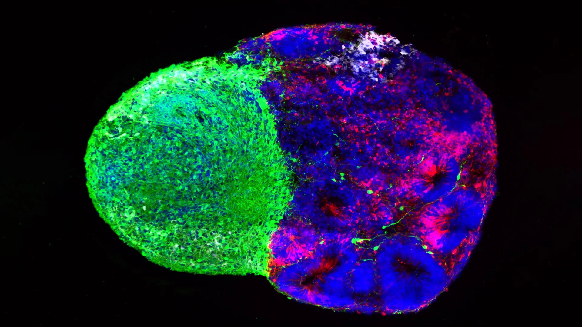
This temporal discrepancy prompted a critical question: what is the functional significance of these premature thalamic projections? Employing advanced human stem cell-derived brain "assembloids," a sophisticated experimental model, the UCLA researchers uncovered a crucial part of the answer. They discovered that these early thalamic projections physically make contact with radial glia during the formative stages of brain development. This physical interaction was found to directly alter the behavior of the radial glial progenitor cells, prompting them to generate a greater number of excitatory neurons, which are the primary signal-carrying neurons within the cortex. This effect was particularly pronounced for upper-layer neurons, a class of neurons that are notably more abundant and contribute to the enhanced complexity of the human brain. Dr. Bhaduri underscores the novelty of this finding, noting, "We already knew that these projections influence how the cortex develops. What we specifically found is that this influence comes through an actual physical connection between the projections and the radial glia — a point of contact that just hasn’t been identified before, and one that very likely does not exist in rodents."
Adding another layer of complexity, the researchers established a connection between this physical interaction and the gene NRXN1, a gene already recognized for its indispensable role in facilitating the formation of neuronal connections. Mutations in NRXN1 have been previously linked to autism spectrum disorder, highlighting a potential molecular bridge between fundamental developmental processes and neurodevelopmental conditions. To further explore this link, the team generated assembloids using cells derived from patients carrying an NRXN1 mutation. In these experimental models, the altered thalamic signals exhibited distinct behavior compared to signals originating from unaffected cells. These modifications in signal transduction consequently altered the delicate balance between the self-renewal of stem cells and the generation of new neurons. This discovery provides a promising avenue for researchers to investigate how disruptions early in brain development, potentially mediated by genetic factors like NRXN1, can influence the intricate organization and functional capacity of the developing cortex.
While these two studies investigated disparate mechanisms—one focusing on cellular metabolism and the other on neural connectivity—they converge on a unifying principle: the developing brain is in a state of constant, dynamic communication. Radial glia, the fundamental building blocks of the cortex, do not operate in isolation. Their critical developmental decisions are continuously shaped and modulated by the complex interplay of signals originating from their immediate microenvironment.
Furthermore, these studies underscore the transformative impact of organoid technology on the field of human brain development research. Just a decade ago, direct experimental investigation into the behavior of uniquely human neural stem cells was severely limited by a lack of appropriate research tools. Today, sophisticated brain organoids and related in vitro models allow scientists to meticulously recreate and study key aspects of human brain development in a controlled laboratory setting. These advanced systems also empower researchers to address fundamental questions that cannot be adequately investigated using traditional animal models alone, thereby accelerating the pace of discovery.
Dr. Bhaduri expresses her hope that these findings will inspire a paradigm shift in how scientists perceive the roles of metabolism and physical cellular interactions in development, encouraging their consideration as active drivers rather than passive background processes. In her words, "Ultimately, these studies give us a glimpse under the hood of how these cells make decisions. Understanding those decisions is a first step toward understanding normal brain development, disease vulnerability and, potentially, how similar stem-cell programs operate in brain cancer." This deeper understanding of cellular decision-making promises to unlock new strategies for addressing neurodevelopmental disorders, neurodegenerative diseases, and even certain forms of cancer.
Related posts:

Related News