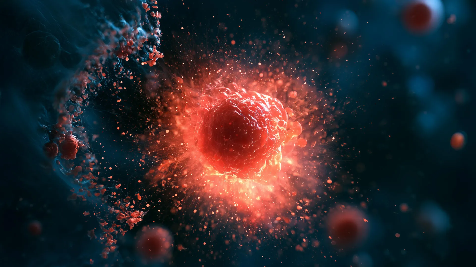
A groundbreaking discovery by Stanford University scientists has illuminated a previously uncharacterized type of immune cell, dubbed "ruptoblasts," which possess a unique and dramatic mechanism for defending the organism: they self-destruct by exploding, effectively acting as microscopic biological munitions. This astonishing cellular event, observed in the humble planarian flatworm, unfolds with such astonishing speed and totality that the entire cell is annihilated and vanishes within mere minutes, leaving behind minimal residual evidence. The research, published in the esteemed journal Cell, delves into the intricate workings of these cells and their potential implications for understanding fundamental biological defense strategies and their potential applications in human medicine.
The genesis of this discovery lies in the extraordinary regenerative capabilities of planarian flatworms, small, unassuming aquatic invertebrates renowned for their remarkable ability to reconstitute their entire bodies from even the smallest fragments. This ancient lineage, having navigated evolutionary pressures for hundreds of millions of years, offers a rich, albeit often overlooked, biological laboratory for exploring fundamental processes like immunity and regeneration. By studying the adaptations that have enabled these creatures to survive and thrive, researchers hope to glean insights that could significantly advance modern therapeutic approaches.
The investigation into these explosive cells was initiated by Chew Chai, a postdoctoral researcher in the laboratory of Bo Wang, an associate professor of bioengineering at Stanford. Chai was initially focused on a long-standing biological enigma concerning flatworms: their capacity to differentiate between self-tissue and foreign tissue. To address this, Chai embarked on a series of experiments involving the surgical fusion of different flatworm individuals. While flatworms are masters of self-repair, the fused "Frankenstein" specimens exhibited a clear rejection response when grafted with tissue from unrelated worms, a phenomenon that bears a striking, albeit superficial, resemblance to the immune rejection of transplanted organs in humans.
However, the cellular choreography underlying this rejection in flatworms proved to be drastically different from anything previously documented in mammalian systems. Chai described the observed reaction as an "enormous inflammatory cascade," akin to an alarm sounding and cells erupting in a violent, explosive manner. This dramatic cellular response immediately signaled the presence of an unconventional defensive mechanism.
Previous investigations into planarian regeneration had already highlighted the critical role of a hormone known as activin. This hormone appears to play a pivotal role in maintaining the delicate balance of the flatworm’s life cycle, with elevated levels potentially impairing regenerative capacity and diminished levels hindering reproductive processes. It was within this context that Chai observed a significant surge in activin levels correlating with the onset of the tissue rejection response in the fused worms. While the animals did not succumb immediately, they perished within days of the fusion. Furthermore, Chai discovered that introducing activin into healthy, unfused flatworms elicited a similar, albeit less severe, inflammatory response, strongly suggesting a direct link between activin signaling and the activation of these peculiar defensive cells.
To dissect the cellular events in real-time, Chai employed advanced techniques such as live cell microscopy and flow cytometry, a sophisticated method utilizing lasers to analyze and sort individual cells. By labeling cells with fluorescent dyes, Chai was able to meticulously track and isolate those that responded to the presence of activin. It was during these high-resolution observations that the explosive nature of the "ruptoblasts" became undeniable. A small population of these cells, upon activation, underwent a rapid and complete disintegration, expelling their cellular contents and annihilating neighboring cells in the process. This entire dramatic sequence, from activation to cellular obliteration, was observed to occur within a remarkably short five-minute window. It was this characteristic explosive destruction that led Chai and Wang to coin the term "ruptosis" to describe the phenomenon and "ruptoblasts" for the cells themselves.
The defining characteristic of ruptosis, setting it apart from other known forms of programmed cell death, is its extraordinary speed and totality. While some mammalian cells and bacteria have been observed to exhibit forms of explosive cell death, these processes typically occur over much longer timescales, often spanning several hours as cellular components are gradually released. In stark contrast, ruptosis is a near-instantaneous event, concluding within seconds to minutes. This rapid discharge of cellular contents transforms each ruptoblast into a highly focused, potent weapon, delivering its payload almost instantaneously rather than through a slow, prolonged leakage.
The efficacy of these explosive cells as defensive agents was further validated through a series of experimental tests. Researchers exposed ruptoblasts to a variety of targets, including the common bacterium Escherichia coli, human kidney cells, and mouse blood cells. In each instance, the ruptoblasts successfully destroyed all tested targets. Crucially, the destructive impact remained precisely localized to the cells in immediate proximity to the ruptoblast explosion. There was no evidence of a propagating chain reaction or the lingering presence of persistent toxicity in the surrounding environment. This ability to deliver a powerful yet highly contained assault suggests significant therapeutic potential, particularly in contexts where targeted destruction of harmful cells is paramount, such as in combating antibiotic-resistant bacterial infections or eradicating tumor cells.
Ruptoblasts also diverge significantly from well-established immune cells like T cells and neutrophils, which are hematopoietic in origin, meaning they are derived from blood stem cells in the bone marrow. Ruptoblasts, in contrast, are classified as glandular cells, indicating a different developmental lineage and potentially a distinct functional role within the immune system. The researchers hypothesize that ruptoblasts achieve their explosive capabilities by dramatically amplifying their normal secretory mechanisms, enabling the sudden and violent expulsion of toxic substances upon encountering activin. A rapid influx of calcium ions from the endoplasmic reticulum within the cell appears to be a critical trigger for initiating and driving the ruptosis process.
The evolutionary history of ruptoblasts further deepens their significance. When Chai extended her search for similar cellular mechanisms in other species, she found them exclusively in basal bilaterians, a group that includes flatworms. This restricted distribution strongly suggests that ruptoblasts represent an ancient immune strategy that emerged early in the evolutionary trajectory of animal life. Chai posits that vertebrates may have lost this particular defense mechanism because their less efficient tissue repair capabilities might render them vulnerable to the collateral damage inflicted by ruptosis. Flatworms, with their abundant stem cells and exceptional regenerative prowess, are far better equipped to compensate for such localized destruction.
The discovery underscores the vastness of unknown biological mechanisms that remain to be uncovered, particularly within organisms that have not traditionally been the focus of mainstream scientific inquiry. "It demonstrates that there are many different immune mechanisms out there," remarked Bo Wang, highlighting the potential for learning from the diverse strategies employed by organisms that inhabit environments teeming with bacteria and viruses. While flatworms may appear simple, their unique biological adaptations could hold the key to novel immune strategies entirely absent in humans and other vertebrates. Wang emphasizes that expanding research beyond traditional model organisms could catalyze innovative approaches to some of medicine’s most intractable challenges.
The research was supported by a multitude of funding sources, including a National Science Foundation Graduate Research Fellowship, a Stanford Graduate Fellowship, a Stanford DARE fellowship, a Human Frontier Science Program grant, a National Institutes of Health grant, and the European Research Council. The study also benefited from the contributions of several other Stanford researchers, including postdoctoral scholar Souradeep Sarkar, former Undergraduate Visiting Research Program scholar Lihan Zhong, Dania Nanes Sarfati, Christine Jacobs-Wagner (a professor in the School of Humanities and Sciences and the School of Medicine), and Hawa Racine Thiam (an assistant professor in the schools of Engineering and Medicine). Additional co-authors, including co-senior author Benyamin Rosental, were affiliated with Ben Gurion University of the Negev. Several of the Stanford researchers hold affiliations with interdisciplinary initiatives such as Stanford Bio-X, the Wu Tsai Neurosciences Institute, and the Maternal & Child Health Research Institute (MCHRI).
About the Author