The relentless progression of neurodegenerative conditions, particularly Alzheimer’s disease (AD), represents a profound global health challenge, impacting millions and placing immense burdens on healthcare systems and families. A central hallmark in the pathology of AD and a spectrum of related disorders, collectively known as tauopathies, involves the dysfunction and aggregation of a crucial protein called tau. Under healthy physiological conditions, tau plays an indispensable role in maintaining the structural integrity of neurons, acting as a stabilizing agent for microtubules—the microscopic, filament-like scaffolding that provides shape, facilitates intracellular transport, and supports the overall functionality of nerve cells. However, in diseased states, this normally beneficial protein undergoes a detrimental transformation, twisting into abnormal, insoluble clumps referred to as neurofibrillary tangles, which severely disrupt brain circuitry and contribute significantly to cognitive decline and neuronal demise.
Recent groundbreaking research conducted by scientists at Sanford Burnham Prebys, detailed in a study published on July 17, 2026, in the esteemed journal Science Advances, sheds new light on a naturally occurring protein that appears to offer significant protection against this tau-induced damage. These findings open promising avenues for the development of innovative therapeutic strategies aimed at bolstering the brain’s intrinsic defensive mechanisms, thereby mitigating the devastating impact of tau-related neurodegeneration.
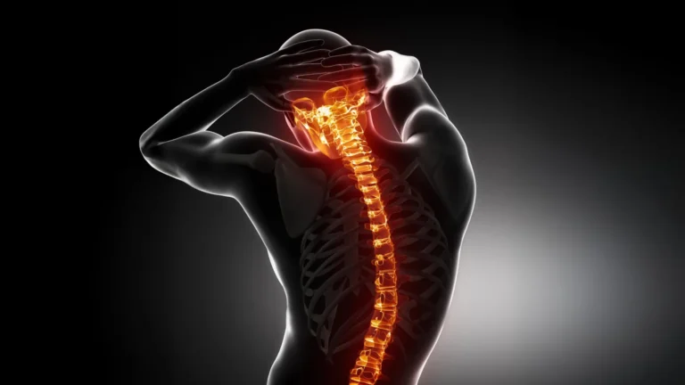
Tau protein is ubiquitous throughout the central nervous system, where its primary function involves supporting the intricate architecture of neurons and the vast interconnected networks they form. This structural support is critical for the efficient transmission of electrical and chemical signals that underpin all cognitive functions, motor control, and sensory processing. In conditions like AD, progressive supranuclear palsy, and frontotemporal dementia, a pathological cascade is initiated where tau proteins detach from microtubules and undergo excessive phosphorylation—a process known as hyperphosphorylation. This over-addition of phosphate groups alters tau’s conformation, causing it to misfold and aggregate into the aforementioned neurofibrillary tangles within the cytoplasm of nerve cells. These abnormal accumulations are directly correlated with synaptic dysfunction, impaired neuronal communication, and ultimately, the death of brain cells, leading to the severe cognitive deficits characteristic of these diseases.
For the past two decades, significant scientific inquiry has focused on understanding the mechanisms behind AD, with considerable attention paid to amyloid-beta (Aβ) plaques, another hallmark pathology. Dr. Timothy Huang, an assistant professor in the Center for Neurologic Diseases at Sanford Burnham Prebys, noted that extensive research from his team and others had previously demonstrated the ability of a protein known as sorting-related receptor with A-type repeats, or SORLA, to suppress the generation and accumulation of amyloid-beta. This established role of SORLA in the amyloid pathway provided a compelling rationale for investigating its potential involvement in the tau pathway, which, despite its critical importance in AD progression, had remained largely unexplored in relation to SORLA. "Comparatively little was understood," Dr. Huang explained, "about whether SORLA exerted any influence on the tau tangles, representing the other primary pathological characteristic observed in Alzheimer’s disease."
To systematically explore SORLA’s potential protective capacity against tau pathology, the research team designed a sophisticated experimental model. They meticulously crossbred mice genetically engineered to produce elevated levels of human SORLA with another strain of mice predisposed to spontaneously develop tau tangles, exhibit brain atrophy, and manifest cognitive impairments—phenotypes highly analogous to human tauopathies. This specific combined model allowed the investigators to precisely examine whether an augmented presence of SORLA could modulate the accumulation of pathological tau and ameliorate the subsequent neurological damage.
The experimental outcomes were highly compelling and provided clear evidence of SORLA’s neuroprotective role. Elevated levels of SORLA were found to interfere significantly with multiple crucial processes implicated in the formation of tau tangles and the progression of neurodegeneration. Specifically, SORLA actively reduced the aberrant hyperphosphorylation of tau, preventing the protein from detaching from microtubules and initiating the misfolding process. Furthermore, SORLA demonstrated the ability to limit the capacity of already misformed tau proteins to act as "seeds"—a critical step in the templated aggregation where abnormal tau recruits healthy tau proteins, leading to the rapid proliferation and growth of larger, more toxic clumps. This dual action on both the initiation and propagation of tau pathology underscored SORLA’s comprehensive protective function.
The beneficial effects observed extended beyond direct modulation of tau protein itself. Mice exhibiting higher SORLA levels maintained healthier synapses—the specialized junctions where neurons communicate with one another. This preservation of synaptic integrity is paramount, as synaptic dysfunction is an early and critical event in AD and other dementias, directly correlating with cognitive decline. Moreover, these mice displayed improved synaptic plasticity, which is the brain’s remarkable ability to strengthen or adjust these connections in response to new experiences or learning. This adaptability is fundamental to memory formation and learning. Dr. Huijie Huang, a staff scientist in the Huang lab at Sanford Burnham Prebys and the lead author of the study, summarized the findings, stating, "When SORLA expression is enhanced, we observed a suppression of the detrimental effects typically found in tauopathies. We were particularly encouraged to find substantial reductions in both brain atrophy and the accumulation of tau, which represents a significant step forward."
To further delineate SORLA’s role, the researchers also investigated the consequences of its absence. It is known that some individuals carry mutations in the Sorl1 gene, which encodes instructions for SORLA production, leading to reduced or dysfunctional SORLA protein. By studying mice genetically modified to completely lack the Sorl1 gene, the team could directly compare the effects of SORLA overexpression with its complete deficiency. As anticipated, these animals experienced the inverse outcome. "Conversely," stated Dr. Tim Huang, the senior and corresponding author of the manuscript, "when the capacity to produce SORLA proteins was abrogated, the harmful effects characteristic of tauopathies were considerably exacerbated." This finding powerfully reinforced the notion that SORLA is not merely a bystander but an active and crucial player in the brain’s defense against tau-mediated neurodegeneration.
To gain a deeper understanding of the molecular mechanisms underlying SORLA’s differential effects based on its abundance, the research team employed an array of advanced sequencing and mapping technologies. These sophisticated methodologies enabled precise measurements of protein levels and gene activity within individual cells, while also providing spatial information regarding the localization of RNA and proteins within complex brain tissue. This granular analysis revealed that an increase in SORLA levels prevented deleterious alterations in protein synthesis at synaptic sites, thereby preserving critical components necessary for neuronal communication. Furthermore, SORLA was found to suppress several other intricate biological pathways known to be associated with the progression of tauopathy, suggesting a broad regulatory influence.
A particularly significant revelation from this analysis involved glial cells. These non-neuronal cells, including astrocytes, microglia, and oligodendrocytes, perform a multitude of essential functions in the brain, ranging from providing structural and metabolic support to neurons, maintaining the brain’s homeostatic environment, and orchestrating immune responses to injury or disease. The study demonstrated that higher SORLA levels attenuated disease-related patterns of gene activity within these glial cells. This modulation of glial cell function is highly relevant, as chronic neuroinflammation mediated by activated glial cells is increasingly recognized as a critical driver of neurodegenerative disease progression. Dr. Huijie Huang highlighted one specific observation, noting, "A particularly notable discovery that provides a foundation for future investigations is the elevated expression of a member of the plexin-B family of receptors in the absence of SORLA." Building on this, Dr. Tim Huang added, "There are existing pharmacological agents designed to target this specific class of receptors, which we might potentially repurpose for the treatment of tau-related dementia disorders. One promising future direction involves exploring the application of these drugs to counteract the overactivation of glial cells, potentially reversing some of the pathological phenotypes observed in tauopathies."
Looking ahead, the research team is focused on meticulously examining how individual types of brain cells respond when SORLA levels fluctuate. Their future experimental designs include grafting human neurons or glial cells into mouse brains, creating "humanized" disease models. This innovative approach will allow them to study the precise effects of different SORLA mutations within a living disease environment that more closely mimics human pathology. "There are inherent differences between mouse and human cells," Dr. Tim Huang explained. "Given that our ultimate goal is to address human disease, it is far more informative to directly observe the modulation and dysfunction of SORLA within the context of human cells situated in a diseased brain."
These future investigations are poised to further elucidate the precise molecular and cellular mechanisms through which SORLA confers protection against toxic tau tangles and to determine whether this natural protective capacity can be therapeutically enhanced. The broader implications of this work extend to identifying existing pharmacological compounds that could be repurposed for the treatment of Alzheimer’s disease and other neurodegenerative conditions primarily driven by tau pathology, offering a potential expedited path to clinical intervention.
This pioneering research was made possible through the dedicated efforts of a large collaborative team, including Christina Huan Shi, Wenqi Yang, Juan C. Piña-Crespo, Jay Bhatnagar, Julian Curatolo, Rabi Murad, Palak Shah, Alex Campos, Alexandra Houser, Rebecca A. Porritt, Giau Van Vo, Tongmei Zhang, Shengjie Feng, and Kevin Y. Yip at Sanford Burnham Prebys, along with Qiang Xiao from The Scripps Research Institute. The study received vital financial support from the National Institutes of Health, specifically the National Cancer Institute and the National Institute on Aging.
About the Author