
A groundbreaking investigation conducted by neuroscientists at Trinity College Dublin has fundamentally reshaped our understanding of early human cognition, demonstrating that infants as young as two months old possess the remarkable capacity to organize visual information into distinct object categories. This sophisticated ability, previously believed to emerge much later in development, suggests that the foundational mechanisms of perception are operational almost from the very inception of life. The revelation offers unprecedented insights into the intricate workings of the infant mind, long before the advent of verbal communication or purposeful motor control.
The findings, recently peer-reviewed and published in the prestigious journal Nature Neuroscience, represent a significant milestone in developmental neuroscience. A collaborative effort from the Trinity College Institute of Neuroscience (TCIN) and the School of Psychology spearheaded this innovative research, which meticulously combined advanced brain imaging techniques with cutting-edge artificial intelligence models to decipher the complex processes occurring within an infant’s brain during its earliest months. This fusion of methodologies provided a unique window into how babies perceive and learn from their surrounding environment.
For centuries, the human mind at birth has been a subject of profound philosophical and scientific debate. Early philosophical perspectives, such as John Locke’s "tabula rasa" concept, posited that the mind begins as a blank slate, with all knowledge and understanding acquired purely through sensory experience. Later developmental psychologists, notably Jean Piaget, outlined a series of cognitive stages, suggesting that complex abstract thought, including object categorization, developed gradually over time, often much later in infancy. This new research from Trinity College Dublin directly challenges these historical assumptions, pushing back the timeline for the emergence of such fundamental cognitive abilities and highlighting a previously underestimated richness in neonatal brain function.
Dr. Cliona O’Doherty, the lead author of the study who conducted this pivotal work while at Trinity’s Cusack Lab, articulated the profound implications of these discoveries. "Parents and researchers alike have long pondered the inner world of an infant, questioning what they genuinely perceive when observing their surroundings. This study vividly illustrates the depth and complexity of brain activity present within the first year of human life," she explained. "Despite the limited communication capabilities of two-month-olds dueostensibly due to their nascent language skills and developing fine motor control, their minds are already engaged in sophisticated processes—not merely registering how things appear, but actively assigning them to specific categories. This unequivocally demonstrates that the core elements of visual cognition are established remarkably early, far sooner than previously anticipated."
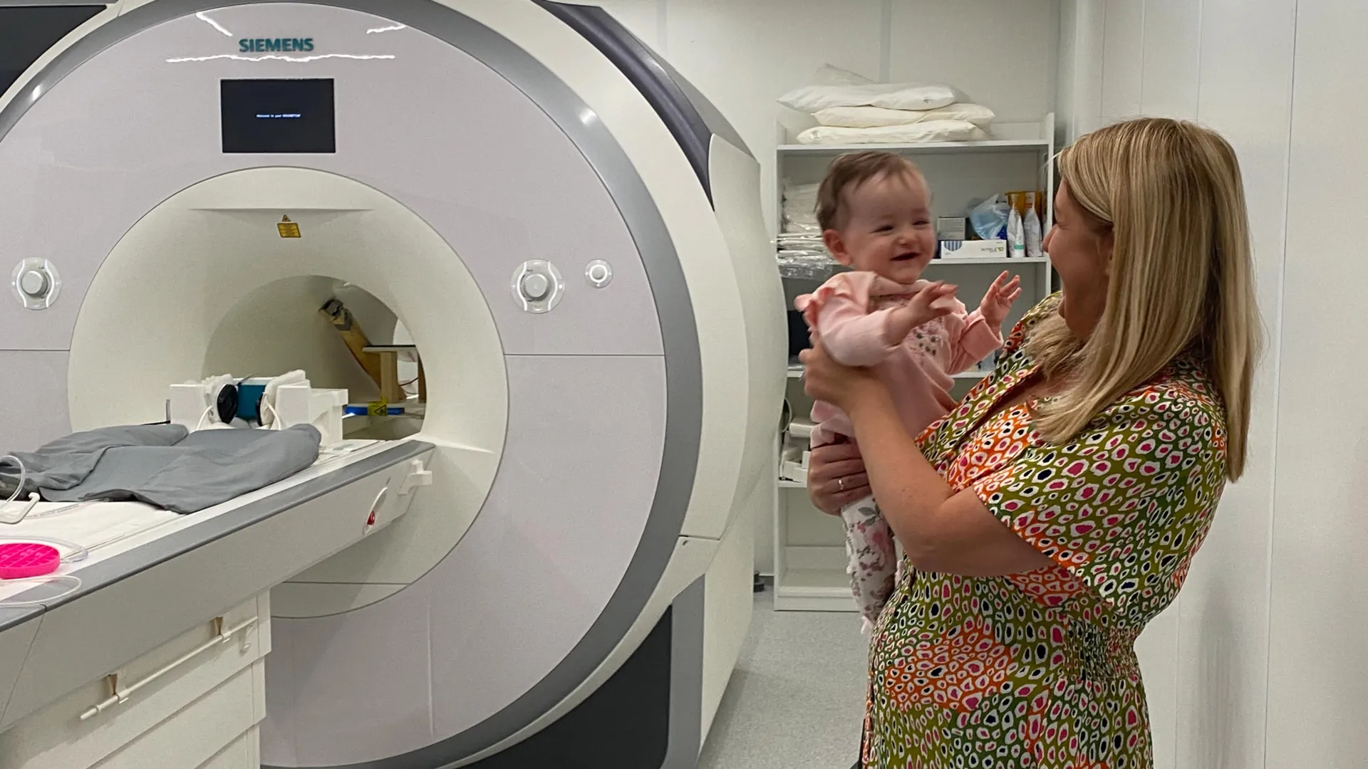
The meticulous execution of this study involved a substantial cohort of participants and an innovative methodological approach. In collaboration with Dublin’s Coombe and Rotunda Hospitals, the dedicated FOUNDCOG research team successfully recruited 130 infants, all precisely two months of age. A critical aspect of the experimental design was ensuring the comfort and engagement of these very young subjects. Each baby was carefully positioned on a soft beanbag, outfitted with specially designed sound-canceling headphones, and presented with a sequence of bright, highly colorful images. These visual stimuli were specifically curated to capture and sustain their attention for periods ranging from 15 to 20 minutes, a crucial duration for data acquisition.
This carefully orchestrated setup facilitated the use of functional Magnetic Resonance Imaging (fMRI), a non-invasive neuroimaging technique that measures brain activity by detecting changes associated with blood flow. When neurons in a particular brain region are active, they require more oxygenated blood, and fMRI can detect these subtle changes. By recording patterns of brain activity as the infants viewed images from 12 distinct and familiar visual categories—such as representations of cats, birds, rubber ducks, shopping carts, and trees—the researchers were able to map the neural responses to specific visual inputs. The ability to perform fMRI on awake infants represents a significant technical achievement, as the procedure typically requires subjects to remain perfectly still, a considerable challenge with newborns. The success of this large-scale longitudinal study on awake infants is a testament to the team’s ingenuity and meticulous planning.
Following the acquisition of this extensive dataset of brain scans, the research team leveraged the power of artificial intelligence to unravel the intricate neural representations. Sophisticated computational models were deployed to analyze how different visual categories were encoded and organized within the infant brain. By systematically comparing the patterns of neural activity observed in the infants’ brains with those predicted by the AI models along established visual recognition pathways, the researchers gained an unprecedented understanding of the mechanisms underpinning early categorization. This synergistic approach, integrating detailed biological data with advanced computational analysis, was instrumental in decoding the complex cognitive processes at play.
Professor Rhodri Cusack, the Thomas Mitchell Professor of Cognitive Neuroscience at Trinity’s School of Psychology and Trinity College Institute of Neuroscience, and a leader of the research team, underscored the monumental scale and potential of this work. "This investigation stands as the largest longitudinal study ever conducted using functional magnetic resonance imaging with awake infants," Professor Cusack stated. "The sheer volume and richness of the captured brain activity data introduce an entirely novel paradigm for assessing the thoughts and perceptions of babies at an exceptionally early developmental stage. Furthermore, it highlights the immense potential for combining neuroimaging with computational models to serve as a powerful diagnostic instrument for very young infants."
Beyond its immediate implications for understanding human development, the study also casts a fascinating light on the future of artificial intelligence. Professor Cusack elaborated on this intriguing connection: "Infants demonstrate a remarkable capacity for learning, often far surpassing the efficiency of contemporary AI models. By meticulously studying the biological mechanisms through which babies acquire knowledge, we aspire to inspire the creation of a new generation of AI algorithms that learn more effectively and with greater efficiency, thereby mitigating their economic and environmental footprint." This vision suggests a future where AI development is informed by the elegant, energy-efficient learning strategies observed in the human brain.
Dr. Anna Truzzi, now a Senior Lecturer in the School of Psychology at Queen’s University Belfast and a co-author of the paper, emphasized the critical role of recent technological advancements in making this research feasible. "Until very recently, reliably measuring how specific regions of an infant’s brain interpreted visual information was beyond our capabilities," Dr. Truzzi noted. "By thoughtfully integrating artificial intelligence with neuroimaging techniques, our study offers a truly unique perspective, significantly deepening our comprehension of how babies learn during their formative first year of life."
The first year is universally recognized as a period of extraordinarily rapid and intricate brain development, laying the groundwork for all subsequent cognitive functions. The new foundational knowledge generated by this study holds profound implications that extend far beyond the laboratory. Dr. Truzzi highlighted several key areas: "This research will be instrumental in guiding early-years educational strategies, informing the development of clinical support for a spectrum of neurodevelopmental conditions, and stimulating the creation of more biologically-grounded methodologies in artificial intelligence."
Professor Eleanor Molloy, a distinguished neonatologist at Children’s Health Ireland and another co-author, stressed the broader societal significance of these findings. "There is an urgent and critical need to enhance our understanding of how neurodevelopmental disorders impact early brain development," Professor Molloy asserted. "The application of awake fMRI, as demonstrated in this study, presents a considerable potential avenue for addressing this pressing challenge." The ability to identify early markers of conditions such as autism spectrum disorder or ADHD could revolutionize diagnostic timelines and enable interventions during the most crucial periods of brain plasticity, potentially altering developmental trajectories for countless children.
The journey of this research, from its inception at Trinity College Dublin to its publication, underscores the collaborative and interdisciplinary nature of modern scientific inquiry. The ongoing work of the researchers, with Dr. O’Doherty now at Stanford University and Dr. Truzzi at Queen’s University Belfast, continues to advance our understanding of the infant mind. Further testament to the profound impact of this research is the artistic interpretation it inspired: acclaimed artist Cian McLoughlin produced original artwork during his tenure as Artist in Residence at the Trinity College Institute of Neuroscience in 2024, reflecting the beauty and complexity of these scientific revelations. This blend of science and art further illuminates the human endeavor to comprehend the earliest sparks of consciousness.
About the Author


