
A groundbreaking advancement in medical diagnostics, developed by a collaborative team from Helmholtz Munich and the Technical University of Munich (TUM), promises to revolutionize the early detection of cardiovascular disease by visualizing the intricate workings of the body’s smallest blood vessels. This innovative imaging modality, dubbed "fast-RSOM" (Raster Scan Optoacoustic Mesoscopy), allows for unparalleled, non-invasive scrutiny of capillary-level vascular health directly through the skin, offering a potential paradigm shift in proactive heart disease management. The technology’s capacity to identify subtle physiological alterations long before the onset of overt symptoms could empower clinicians with crucial lead time, enabling more personalized therapeutic strategies and fostering improved long-term cardiovascular well-being.
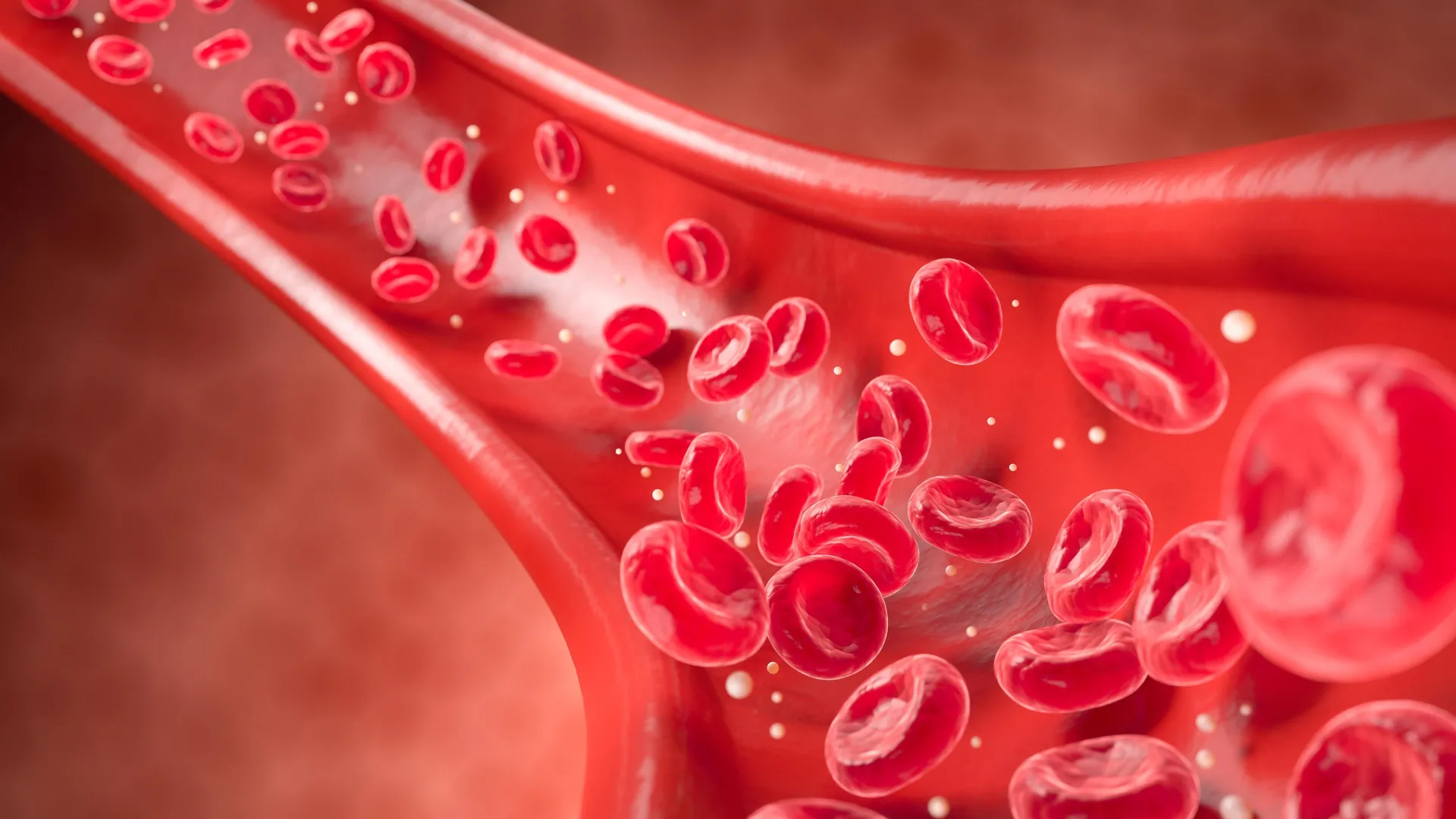
Cardiovascular ailments, in their nascent stages, often betray their presence through insidious changes within the body’s most diminutive vascular networks. These initial disruptions manifest as subtle impairments in the blood vessels’ ability to appropriately dilate and constrict, a condition medically termed microvascular endothelial dysfunction (MiVED). Historically, the precise, non-invasive observation and quantification of these microvascular anomalies in living individuals has remained an elusive goal, posing a significant hurdle to early intervention.
"For the first time, fast-RSOM affords us the capability to non-invasively evaluate endothelial dysfunction at the resolution of single capillaries and within the skin’s layers in human subjects," stated Dr. Hailong He, the study’s lead author and a researcher affiliated with the Institute of Biological and Medical Imaging at both Helmholtz Munich and TUM. Complementing this sentiment, Dr. Angelos Karlas, a co-first author, who is also a practicing Vascular Surgeon and a Senior Research Scientist at TUM University Hospital, remarked, "Our innovative methodology provides an unprecedented perspective on the granular manifestations of cardiovascular disease at the microvascular stratum."
The fast-RSOM system is engineered to capture high-resolution, dynamic biomarkers intrinsically linked to MiVED, thereby illuminating minute yet significant deficits in blood vessel functionality. These physiological indicators frequently surface considerably in advance of any discernible symptoms or macroscopic indicators of cardiovascular pathology. Such microvascular derangements are commonly correlated with established risk factors, including tobacco use, elevated blood pressure, and obesity.
Instead of relying solely on the presence of these risk factors to estimate an individual’s susceptibility, fast-RSOM directly quantifies the tangible effects that these conditions have already exerted on the microvascular architecture. This direct measurement permits healthcare providers to ascertain the functional status of the smallest blood vessels at a stage where serious complications have yet to emerge. The identification of these prescient signals heralds new avenues for earlier diagnosis, more effective preventative measures, and the more accurate surveillance of cardiovascular health. The technology holds the promise of identifying individuals at elevated risk with enhanced precision and of meticulously tracking the impact of lifestyle modifications or medical interventions on the functional integrity of blood vessels over time.
The research consortium is actively pursuing the validation of fast-RSOM across broader and more heterogeneous patient cohorts, with a clear objective of integrating its unique biomarkers into the routine fabric of clinical practice. The inherent portability, rapid operational speed, and non-invasive nature of the device position it as a potential tool for deployment in outpatient settings, augmenting conventional cardiovascular risk assessment protocols.
"By facilitating earlier therapeutic interventions and more refined monitoring, fast-RSOM possesses the potential to fundamentally reshape the landscape of cardiovascular disease prevention and management, ultimately leading to improved patient outcomes and a reduction in overall healthcare expenditures," posited Professor Vasilis Ntziachristos, Director of the Bioengineering Center at Helmholtz Munich and a distinguished Professor of Biological Imaging at TUM.
Raster Scan Optoacoustic Mesoscopy (RSOM) itself represents a sophisticated non-invasive imaging methodology that leverages transient light pulses to elicit ultrasound signals. This process generates exceptionally detailed three-dimensional renderings of subcutaneous structures. Its remarkable sensitivity allows for the detection of minute alterations in blood vessels, oxygen saturation levels, and tissue composition that often elude detection by conventional imaging modalities. The synergy of strong intrinsic contrast with the ability to penetrate to a significant depth makes RSOM an invaluable asset for the early identification of conditions such as cardiovascular disease and diabetes. Furthermore, its inherently compact design suggests the potential for wider accessibility of advanced diagnostic capabilities beyond the confines of specialized research institutions. The foundational development of this technology is credited to the research group led by Professor Vasilis Ntziachristos.
Dr. Hailong He, a key figure in this research, is a dedicated researcher contributing to the fields of biological and medical imaging at both Helmholtz Munich and the Technical University of Munich (TUM). His work is integral to advancing our understanding of the body’s intricate biological processes through innovative imaging techniques.
Dr. Angelos Karlas, another principal contributor, is a board-certified Vascular Surgeon in Germany and serves as a Senior Research Scientist within the Clinic and Polyclinic for Vascular and Endovascular Surgery at the TUM University Hospital, Rechts der Isar, located in Munich. His clinical expertise is complemented by his research leadership role in clinical studies at the Chair for Computer-Aided Medical Procedures and Augmented Reality at TUM.
Professor Vasilis Ntziachristos, a pivotal leader in this endeavor, holds significant academic and directorial positions. He serves as the Director of the Bioengineering Center and the Director of the Institute of Biological and Medical Imaging at Helmholtz Munich. Concurrently, he chairs the Biological Imaging program at the Technical University of Munich (TUM). He is also a founding member and an active participant on the Board of Directors of TranslaTUM, TUM’s Central Institute for Translational Cancer Research. Additionally, he maintains an affiliation with the Munich site of the German Centre for Cardiovascular Research (DZHK), underscoring his broad impact on cardiovascular research.
About the Author


