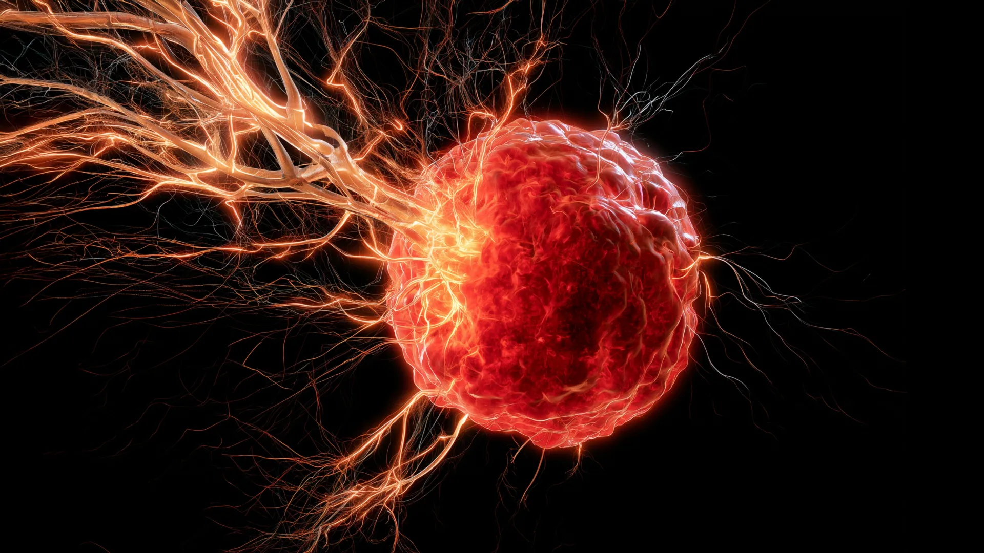
A groundbreaking investigation conducted at the University of Oklahoma has illuminated a sophisticated interplay between aggressive breast cancer cells and the body’s own defense mechanisms, revealing how tumors can actively enlist the nervous system to foster their own proliferation and potentially evade therapeutic interventions. This research delves into a previously obscured pathway by which cancerous growths in certain breast cancer subtypes establish a robust neural infrastructure within their confines, a development that may significantly influence disease progression and treatment efficacy.
For an extended period, the medical and scientific communities have acknowledged the pervasive presence of intricate nerve networks within solid tumors. However, the precise biological machinations that facilitate the ingress of these neural fibers into the tumorous mass have remained largely enigmatic. The recent findings, disseminated in the esteemed scientific journal Cell Death & Differentiation, offer a compelling explanation for this phenomenon, particularly within the context of triple-negative breast cancer, a formidable and challenging variant of the disease characterized by its aggressive nature and limited targeted treatment options.
At the heart of this revelation is the discovery that tumor microenvironments actively solicit the assistance of specific immune cells, notably macrophages. These versatile cells, integral to the immune system’s frontline defense against pathogens and crucial for orchestrating tissue repair, are drawn into the tumor’s vicinity. Once embedded within the cancerous tissue, these tumor-associated macrophages assume a critical role in the recruitment of neural elements. They achieve this by secreting a potent signaling molecule known as brain-derived neurotrophic factor (BDNF). BDNF, a protein predominantly recognized for its neurotrophic functions—supporting the survival, growth, and differentiation of neurons within the central nervous system—exhibits an unexpected and detrimental behavior within the tumorous landscape.
The research elucidates how cancer cells cunningly exploit this endogenous signaling pathway. By inducing the localized release of BDNF, tumors effectively "call" for nearby nerves to extend their tendrils and infiltrate the cancerous mass. This aberrant neurogenesis within the tumor is hypothesized to contribute significantly to the cancer’s ability to grow unchecked and, crucially, to develop resistance against conventional therapeutic agents. Dr. Maureen Cox, an assistant professor in the Department of Microbiology and Immunology at the OU College of Medicine and a distinguished researcher at the OU Health Stephenson Cancer Center, emphasized the pivotal role of macrophages in this process. "Macrophages are the critical source for drawing nerves into the tumor," Dr. Cox stated. "Although macrophages typically play a positive role in the body, they are facilitating a negative function in this scenario of breast cancer." This observation underscores a concerning subversion of normal biological processes by the malignant cells.
The implications of this intricate neural infiltration extend beyond mere structural support for the tumor; it suggests a potential avenue for novel therapeutic strategies. The conventional paradigm of cancer treatment primarily focuses on directly eliminating malignant cells. However, this research opens the door to an alternative approach: disrupting the communication channels that facilitate tumor growth. By targeting the signaling nexus between macrophages and the nerves they recruit, future therapies might succeed in inhibiting the very infrastructure that appears to bolster tumor progression and treatment resistance.
To rigorously test this therapeutic hypothesis, Dr. Cox and her research team embarked on preclinical investigations utilizing a murine model. Their experiments involved administering a pharmacological agent designed to specifically block BDNF signaling. The results were highly encouraging: the intervention effectively prevented nerve infiltration into the tumors. Concurrently, a significant reduction in tumor growth rates was observed, providing compelling evidence for the therapeutic potential of targeting this newly identified pathway. "It looks really promising that we can use this drug, which is already on the market, to target BDNF," Dr. Cox remarked. She further elaborated on the potential broader impact of this intervention, suggesting, "We believe that the nerves are immunosuppressive, so if we can stop the nerves from growing in the first place, maybe we can boost the immune response to help fight the cancer." This hypothesis implies that the presence of nerves within tumors might actively dampen the anti-cancer immune response, and by removing them, the body’s natural defenses could be reactivated.
Further bolstering the translational relevance of these findings, the researchers extended their analysis to human patient data. They meticulously examined samples from individuals diagnosed with triple-negative breast cancer, seeking to ascertain whether the observed biological mechanisms in the laboratory models were indeed mirrored in human disease. The correlational analysis revealed a significant association: tumors exhibiting elevated levels of both macrophages and BDNF were consistently linked with poorer patient survival outcomes. This evidence strongly suggests that the neural infiltration pathway elucidated in mice is not merely a laboratory artifact but plays a demonstrably important role in the clinical trajectory of triple-negative breast cancer in humans.
The research team remains focused on unraveling the intricate ways in which these intruding nerves contribute to tumor advancement. Current hypotheses suggest several possible mechanisms. One compelling theory is that nerves may actively promote the formation of new blood vessels, a process known as angiogenesis, which is essential for supplying tumors with the oxygen and nutrients required for their rapid proliferation. Another line of inquiry explores whether cancer cells might utilize existing nerve pathways as conduits for their dissemination, facilitating metastasis—the spread of cancer from the primary site to distant parts of the body.
Looking ahead, the researchers are not limiting their investigation to breast cancer. They are planning to extend their studies to high-grade ovarian cancer, another aggressive and therapeutically challenging malignancy that shares certain biological characteristics with triple-negative breast cancer. This comparative approach aims to determine if the identified mechanism of neural recruitment is a common feature across different aggressive cancer types. "Ultimately, we want to turn the anti-tumor immunity back on in cancer patients so their own immune systems can reject the tumors," Dr. Cox articulated her ultimate goal, highlighting the ambition to harness the patient’s innate immune capabilities for cancer eradication.
The scientific endeavor behind this groundbreaking discovery was generously supported by several prestigious funding bodies. Key among them were the National Institute of General Medical Sciences of the NIH, which provided critical funding through award numbers P20GM103447 and P20GM103639. Additional vital support was received from Oklahoma’s Tobacco Settlement Endowment Trust (TSET), a primary benefactor of the Stephenson Cancer Center and the TSET Health Promotion Research Center at the University of Oklahoma. Furthermore, the Oklahoma Shared Clinical and Translational Resources, through an Institutional Development Award from the National Institute of General Medical Sciences (grant no. U54GM104938), also contributed significantly to the successful execution of this research. These collaborative funding efforts underscore the importance and multidisciplinary nature of this investigation into the complex biology of cancer.
About the Author