
Precision Uncovered: Scientists Map the Brain’s Norepinephrine Command Center
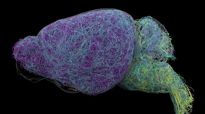
A groundbreaking study has fundamentally reshaped our understanding of how a vital brain region, the locus coeruleus (LC), orchestrates communication within the nervous system. Traditionally viewed as a generalized broadcast station for the neurotransmitter norepinephrine, new research published in Nature reveals that this small cluster of neurons operates with remarkable precision, dispatching targeted messages through distinct, specialized pathways. This paradigm shift, spearheaded by researchers at the Allen Institute, transforms the long-held "loudspeaker" analogy for the LC into one of a highly sophisticated, targeted postal network, with profound implications for treating neurological and psychiatric conditions.
The locus coeruleus, whose name derives from the Latin for "blue place" due to its distinctive pigmentation, is a remarkably compact nucleus nestled deep within the brainstem. Despite containing a mere fraction of the brain’s total neurons, its influence is disproportionately vast. Its primary function revolves around the production and release of norepinephrine (NE), a crucial chemical messenger that plays a central role in a myriad of physiological and cognitive processes. Norepinephrine is instrumental in regulating states of arousal, vigilance, and attention, influencing our heart rate, blood pressure, and the body’s adaptive responses to stress and excitement. Its ubiquitous presence throughout the brain and spinal cord previously led scientists to assume a largely undifferentiated signaling mechanism, where NE signals were thought to be broadly disseminated across wide cerebral areas simultaneously.
This conventional view has now been challenged by an exhaustive investigation employing a powerful combination of cutting-edge methodologies. The Allen Institute team utilized advanced brain imaging techniques to visualize neural circuits, meticulously recorded the electrical activity of individual neurons, applied sophisticated genetic tools to selectively tag and manipulate specific cell populations, and conducted intricate behavioral experiments with mice. These multi-modal approaches allowed for an unprecedented level of detail in mapping the LC’s intricate connectivity and functional dynamics, leading to the discovery that its communication strategy is far more nuanced and organized than previously imagined. Rather than emitting a uniform, global signal, the LC strategically delivers tailored messages to specific neural destinations, with different groups of neurons activating in response to unique experiences and demands.
A cornerstone of this discovery is the revelation of distinct anatomical segregation within the LC, dictating the precise reach of its norepinephrine signals. The researchers painstakingly traced the axonal projections – the long, slender extensions of nerve cells that transmit electrical impulses – from individual LC neurons to their target regions. They found a clear division of labor: neurons situated in the upper, or dorsal, portion of the LC primarily project to higher-order brain regions, including the cerebral cortex and the forebrain. These areas are critically involved in complex cognitive functions such as perception, memory, decision-making, and conscious thought. Conversely, cells located in the lower, or ventral, part of the LC predominantly send their signals downward to the brainstem and spinal cord, regions vital for regulating basic life-sustaining functions, motor control, and sensory processing. This elegant organizational scheme means that different segments of the LC are uniquely positioned to modulate separate, yet interconnected, brain functions, providing a structural basis for its targeted communication.
Karel Svoboda, a director of Neural Dynamics at the Allen Institute and a co-author of the study, eloquently summarized this shift in understanding, stating, "The findings suggest the brain’s norepinephrine system is far more like a targeted postal network than a foghorn." This analogy underscores the precise and compartmentalized nature of NE signaling, moving away from a generalized influence towards a highly specific regulatory role.
The implications of this refined understanding extend significantly to the realm of therapeutic interventions for a spectrum of neurological and mental health disorders. Norepinephrine’s critical role in mood regulation, attention, and cognitive function makes it a prime target for pharmacological treatments for conditions such as major depression, anxiety disorders, and Attention-Deficit/Hyperactivity Disorder (ADHD). Current medications designed to modulate NE levels, such as certain antidepressants and stimulants, often exert their effects broadly across multiple brain circuits. While beneficial in some areas, this lack of specificity can lead to undesirable side effects in others, limiting their efficacy and tolerability. For instance, a drug aimed at improving attention might also inadvertently affect sleep patterns or heart rate due to its widespread action.
Furthermore, LC neurons are among the earliest brain cells to exhibit degeneration in the progression of Alzheimer’s disease, suggesting that disruptions in NE signaling may contribute significantly to the cognitive decline characteristic of this neurodegenerative condition. A detailed "map" of the LC’s specialized pathways, as provided by this research, offers an unprecedented opportunity to develop a new generation of highly selective therapies. By targeting specific NE circuits implicated in particular symptoms or disease processes, scientists could potentially design treatments that precisely address pathological mechanisms while minimizing off-target effects, thereby enhancing both efficacy and patient quality of life. The research received vital support from the National Institutes of Health’s Brain Research Through Advancing Innovative Neurotechnologies® Initiative, known as The BRAIN Initiative®, highlighting its strategic importance in advancing neuroscience.
Beyond anatomical distinctions, the study also uncovered critical functional differences between these segmented LC neuronal populations, particularly in the context of learning and environmental engagement. The researchers observed that neurons within the dorsal LC, those projecting to the cerebral cortex, exhibited heightened activity during learning processes. In behavioral experiments, mice engaged in decision-making tasks were monitored as they adjusted their choices following an unfavorable outcome. It was precisely during these moments of adaptive learning and error correction that the activity of these dorsal LC neurons surged, indicating their direct involvement in cognitive flexibility and the integration of new information.
In stark contrast, neurons located in the ventral LC displayed a distinct pattern of activity. These cells showed an increase in firing rate shortly before mice chose to disregard environmental cues that signaled a potential reward. This suggests that the ventral LC population plays a crucial role in regulating an animal’s overall engagement with its surroundings, influencing whether it responds to or disengages from opportunities present in its environment. Svoboda reiterated this functional mapping: "What emerged was a clear map: neurons in the dorsal LC that send signals upward to the cortex are involved in learning, whereas neurons in the ventral region projecting downward to the brainstem and spinal cord govern whether animals engage with their environment at all." Intriguingly, these anatomical and functional divergences were further substantiated by distinct patterns of gene expression within the two neuronal groups, providing molecular evidence for their specialized roles and identities.
The sheer scale and depth of the investigation itself yielded another remarkable finding concerning the structural characteristics of LC neurons. The study involved an extensive analysis of nearly 35,000 neurons using whole-brain imaging, profiling the genetic makeup of almost 400,000 individual cells, and meticulously reconstructing the complete three-dimensional structures of several exceptionally complex nerve cells. During this painstaking work, researchers discovered that LC neurons possess extraordinarily long axons, the conduits responsible for transmitting neural signals over considerable distances. On average, these axons measured approximately 35 centimeters in length within the mouse brain. One particular axon stood out, boasting an astonishing reconstructed length of 70.32 centimeters, making it part of the longest single neuron ever fully mapped in a mouse.
This remarkable physical characteristic allows LC neurons to span vast regions of the brain, underscoring their capacity for widespread influence. However, despite their extensive reach, these super-long axons maintain a surprising degree of selectivity in their targeting. Jeremiah Cohen, a scientist at the Allen Institute and co-author of the study, commented, "This neuron, like many others that we studied, supplies NE to a very large volume of the cerebral cortex. For the brain, this is highly unusual. Most neurons are more specific in their targets." Yet, he emphasized, "But this neuron doesn’t release NE everywhere. It ignores the cerebellum, brainstem, and spinal cord." This paradox of vast reach coupled with precise targeting further reinforces the study’s central tenet: the LC operates through carefully organized, highly selective signaling pathways, optimizing efficiency by delivering its neuromodulatory influence exactly where and when it is needed.
The research also uncovered intriguing parallels between the norepinephrine system and the brain’s dopamine signaling system, another major neurotransmitter involved in reward, motivation, and motor control. Dopamine pathways are well-known for their projections to the basal ganglia, a brain region critical for habit formation and motor learning. Scientists now hypothesize that dopamine and norepinephrine may work in concert, potentially allowing the brain to acquire and process multiple complex and abstract concepts simultaneously. This collaborative effort between distinct neuromodulatory systems could provide a more comprehensive framework for understanding the intricate mechanisms underlying learning and adaptive behavior.
By meticulously revealing the intricate connections, specialized functions, and unusual physical architecture of neurons within the locus coeruleus, this seminal research provides an unprecedentedly detailed portrait of how the mammalian brain communicates and learns. While these findings emerged from experiments conducted in mice, the fundamental principles of neural organization and signaling are often conserved across species. Therefore, this work holds immense promise for eventually deepening our understanding of human brain function and for guiding the development of novel, highly targeted therapeutic strategies that precisely intervene in specific neural circuits, rather than broadly affecting entire signaling systems, paving the way for more effective and personalized treatments for a range of debilitating brain disorders.

Related News