
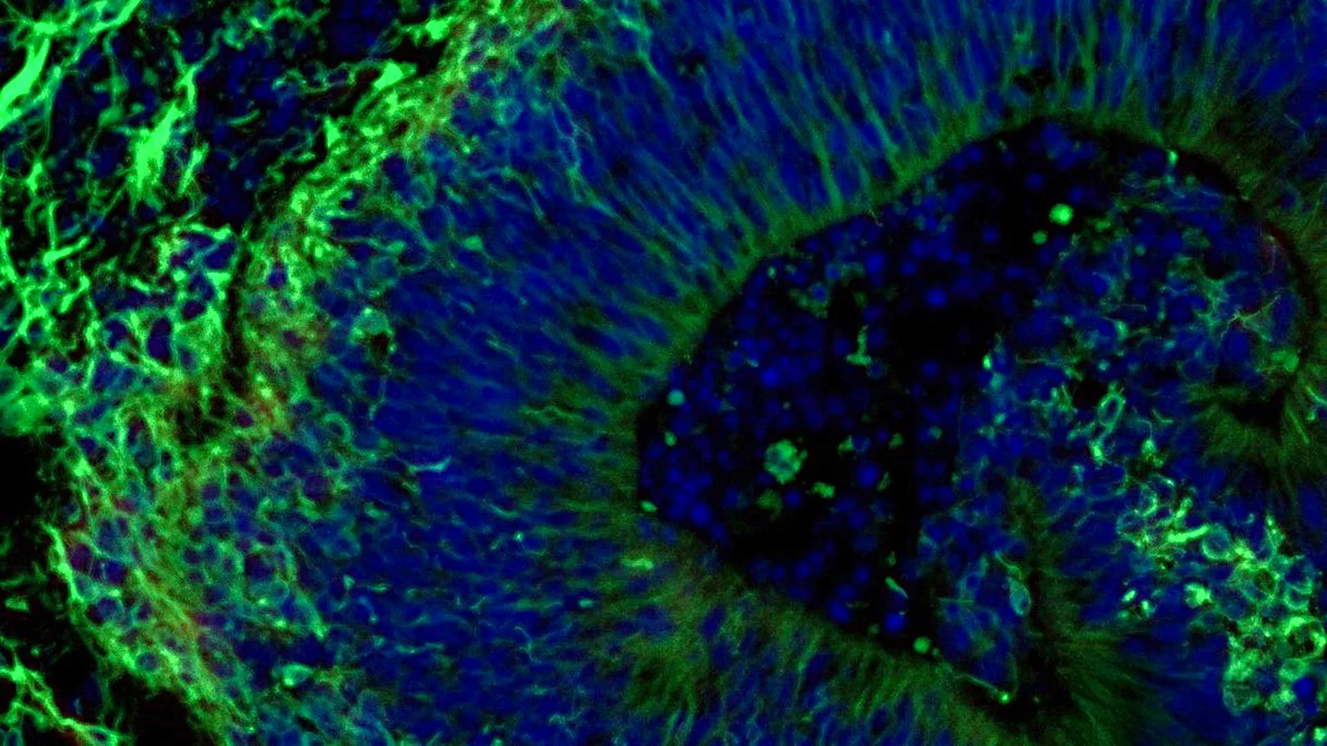
Groundbreaking research conducted at Johns Hopkins Medicine has unveiled compelling evidence suggesting that miniature brain structures, meticulously cultivated from the cellular material of individuals diagnosed with Alzheimer’s disease, possess the remarkable potential to predict a patient’s likely response to specific pharmaceutical interventions aimed at alleviating the complex neuropsychiatric manifestations of the condition. This advancement marks a significant stride toward personalized therapeutic strategies for a disease that currently affects millions and lacks a definitive cure.
The cornerstone of this investigation lies in the utilization of laboratory-created brain tissues, scientifically termed organoids, which serve as sophisticated in vitro models of human brain architecture. These findings contribute to an expanding body of scientific consensus that champions these scaled-down neural constructs as a promising avenue for the development and selection of highly individualized treatment regimens for distinct patient cohorts afflicted by Alzheimer’s disease. Alzheimer’s disease stands as the preeminent cause of dementia globally, impacting a substantial segment of the population, with over seven million individuals in the United States alone grappling with its debilitating effects.
Beyond their predictive capacity for drug efficacy, the research team also identified the release of minute particles, known as extracellular vesicles, from these engineered brain tissues. These vesicles, acting as intercellular couriers, are laden with crucial cellular information, and their analysis may herald the discovery of novel biomarkers. Such biomarkers could prove invaluable for both the early diagnosis of Alzheimer’s disease and the precise determination of its progression stage. This dual potential—predicting treatment outcomes and offering diagnostic insights—underscores the profound implications of this research.
The study, which garnered partial financial support from the National Institutes of Health, has been formally documented and disseminated within the pages of Alzheimer’s & Dementia: The Journal of the Alzheimer’s Association, a highly respected publication in the field of neurodegenerative research. This peer-reviewed publication lends significant credibility to the findings and paves the way for further scientific exploration and validation.
The concept of employing these miniature brain models for the facilitation of personalized care is a central theme emerging from this work. Dr. Vasiliki Machairaki, Ph.D., an associate professor of genetic medicine at the Johns Hopkins University School of Medicine and the principal investigator of the study, articulated the transformative potential of these organoids. She stated, "Our study suggests that large-scale, patient-derived brain organoids and the vesicles they secrete can help us stage Alzheimer’s disease, investigate the mechanisms that drive it and assess how patient subgroups may respond to different treatments." This multifaceted utility highlights the comprehensive nature of the insights these models can provide.
While the quest for a cure for Alzheimer’s disease continues, current therapeutic approaches often focus on managing the constellation of neuropsychiatric symptoms that accompany the disease. These symptoms, which can encompass anxiety, depression, and agitation, are nearly ubiquitous among affected individuals. Selective serotonin reuptake inhibitors (SSRIs), a class of antidepressants, are frequently prescribed to mitigate these distressing manifestations. However, a significant clinical challenge lies in the considerable variability observed in patient responses to these medications, a phenomenon that Dr. Machairaki’s research seeks to unravel.
The Johns Hopkins research team meticulously focused their efforts on constructing and examining miniature models representative of the hindbrain. This specific region of the brain, situated at the posterior of the skull, is critical for regulating fundamental life-sustaining functions, including respiration, sleep-wake cycles, and heart rate regulation. The researchers’ primary objective was to ascertain whether these engineered hindbrain models could reveal distinct molecular signatures that would indicate whether a particular SSRI, specifically escitalopram oxalate, might prove effective in ameliorating the neuropsychiatric symptoms associated with Alzheimer’s disease.
The innovative process of generating these brain organoids commenced with the collection of blood samples, obtained with the explicit consent of individuals participating in Alzheimer’s disease research at the NIH-funded Johns Hopkins Alzheimer’s Disease Research Center. This ethical and consent-driven approach is fundamental to all human subject research.
Following sample collection, the research team employed sophisticated cellular reprogramming techniques to revert the collected blood cells to a pluripotent stem cell-like state. These reprogrammed cells, designated as induced pluripotent stem cells (iPSCs), possess an extraordinary capacity to differentiate into virtually any cell type found within the human body, thus serving as a versatile biological building block.
Leveraging iPSCs derived from both individuals diagnosed with Alzheimer’s disease and healthy control subjects, the researchers meticulously cultivated hindbrain organoids. These organoids were populated with specialized brain cells, known as neurons, specifically those capable of synthesizing the crucial neurotransmitter serotonin. This targeted approach ensured that the organoids recapitulated key neural characteristics relevant to the study’s objectives.
Through a carefully orchestrated process, these iPSCs were guided to self-organize into compact, pea-sized clusters of brain tissue, effectively mimicking the structural organization of the hindbrain. The study’s impressive scale included hundreds of these organoids, each meticulously representing an individual participant, encompassing both Alzheimer’s patients and healthy controls. Dr. Machairaki posited that this extensive collection might represent one of the most substantial brain organoid studies undertaken to date within the Alzheimer’s research landscape.
Analysis of these patient-derived organoids revealed significant and distinct molecular alterations when compared to organoids generated from healthy individuals. These differences were observed at a fundamental molecular level, mirroring key biological characteristics of Alzheimer’s disease. Specifically, the organoids derived from Alzheimer’s patients exhibited discernible variations in proteins implicated in intercellular communication within the brain, inflammatory processes, and pathways directly associated with the disease’s pathogenesis.
Subsequently, the researchers introduced escitalopram oxalate, a widely prescribed antidepressant medication, to these organoids. The impact of this pharmacological intervention was then rigorously assessed at the molecular level.
In a subset of organoids originating from Alzheimer’s patients, the administration of escitalopram led to an observable increase in specific proteins. These proteins were found to be integral to serotonin signaling pathways and the intricate mechanisms of communication between brain cells—precisely the pathways targeted by antidepressant medications. Conversely, other organoids demonstrated a minimal or entirely absent molecular response to the drug.
"We used these organoids to model how some patients’ tissue may respond to a commonly prescribed SSRI," Dr. Machairaki explained, elaborating on the experimental design. "On a large-scale level, our model may eventually be used to identify subgroups of patients, based on underlying molecular mechanisms, who are more likely to respond to certain drugs and thus help us to create precise, targeted treatments in the long run." This statement encapsulates the ultimate aspiration of the research: to translate in vitro findings into tangible clinical benefits.
The investigation then pivoted to exploring the diagnostic and predictive potential of the extracellular vesicles released by the organoids. The researchers sought to determine if these tiny cellular packages could serve as reliable biomarkers for Alzheimer’s disease or as indicators of how neural tissue might respond to therapeutic interventions.
Prior to and following the administration of escitalopram, the scientists meticulously analyzed the protein content within extracellular vesicles secreted by both the patient-derived organoids and the healthy control organoids. This comparative analysis was crucial for identifying disease-specific or treatment-responsive molecular signatures.
The extracellular vesicles were found to be rich in proteins essential for a wide array of brain functions. These included proteins involved in neuronal communication, memory consolidation, and the regulated release of neurotransmitters, underscoring the vesicles’ role as carriers of vital neural information.
A significant observation was the identification of clear alterations in several disease-associated proteins within vesicles derived from organoids of individuals with Alzheimer’s disease. Specifically, proteins such as RAB3A, NSF, and ATCAY, which are critical for the normal signaling processes between brain cells, were found at lower concentrations in the Alzheimer’s organoids.
Intriguingly, after treatment with escitalopram, the levels of certain proteins within these vesicles exhibited an increase in specific samples. These molecular shifts were particularly pronounced in proteins associated with serotonin signaling and synaptic pathways, the very targets of antidepressant medications.
The observed heterogeneity in organoid responses—some displaying robust molecular changes while others showed minimal to no alteration—led Dr. Machairaki to propose a groundbreaking possibility. She suggested that extracellular vesicles derived from brain organoids could, in the future, offer a non-invasive means of identifying which patients are most likely to derive benefit from a particular therapeutic agent.
Looking ahead, Dr. Machairaki has ambitious plans for the evolution of these brain organoid models. She aims to develop more sophisticated organoids that incorporate immune cells and vascular-like networks, thereby more closely mimicking the intricate architecture and functional complexity of living human brain tissue. The inclusion of these cellular components is expected to enhance the physiological relevance and predictive accuracy of the models.
With continued research and refinement, Dr. Machairaki envisions a future where extracellular vesicles could function as a form of "liquid biopsy." Such a non-invasive diagnostic tool would hold the potential to revolutionize the management of Alzheimer’s disease by enabling early detection, accurate staging, and the identification of specific disease subtypes, thereby paving the way for highly tailored treatment strategies.
Dr. Machairaki underscored that the current study represents a foundational step in a long-term research endeavor, acknowledging the complexities and ongoing challenges in the field of Alzheimer’s disease research. The collaborative effort involved a multidisciplinary team, including researchers from Johns Hopkins, the University of Rochester School of Medicine and Dentistry, and Tymora Analytical Operations, all contributing their expertise to this significant undertaking. The research was supported by substantial funding from the National Institutes of Health, the Paul G. Allen Frontiers Foundation, and the Richman Family Precision Medicine Center of Excellence in Alzheimer’s Disease at The Johns Hopkins University, reflecting a concerted effort to advance understanding and treatment of this devastating disease.
About the Author












