Researchers affiliated with The University of Alabama in Huntsville (UAH), a constituent institution of The University of Alabama System, have unveiled a groundbreaking application for continuous, low-intensity ultrasound therapy, potentially revolutionizing the approach to preventing post-traumatic osteoarthritis and accelerating the healing of joint injuries. This novel, non-invasive modality demonstrates an extraordinary capacity to redirect the body’s inherent immune response, steering it away from chronic inflammation and towards a robust tissue repair pathway, thereby presenting a promising, drug-free strategy for significantly improving recovery outcomes.
The cornerstone of this significant scientific advancement lies in a collaborative study, meticulously detailed within the esteemed scientific journal Scientific Reports, published by Nature. At the helm of this interdisciplinary endeavor was Dr. Anuradha Subramanian, a distinguished professor in chemical and materials engineering, whose leadership was instrumental in integrating biological insights derived from the doctoral research of Dr. Shahid Khan with sophisticated computational and statistical analyses meticulously developed by Dr. Satyaki Roy, a professor of mathematical sciences. The project also benefited from the dedicated contributions of graduate student Owen Trippany, and its ambitious scope was made possible through vital funding secured from the National Institutes of Health, specifically via an R01 grant awarded to Dr. Subramanian.
At the heart of this investigation lies a profound exploration into the intricate mechanisms by which ultrasound exerts its influence on critical immune cells known as macrophages. These specialized cellular entities are recognized for their dual roles, acting as both architects of inflammation and facilitators of tissue regeneration. The research team meticulously examined how macrophages, under the influence of continuous low-intensity ultrasound, adapt their functional states.
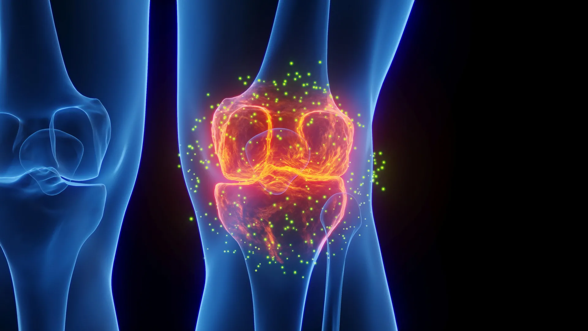
Dr. Subramanian elucidated the complex immunological ballet that unfolds post-injury, explaining, "Following an injury, the body orchestrates an influx of inflammatory ‘defender’ macrophages, designated as M1, whose primary responsibility is the clearance of damaged cellular debris. Concurrently, ‘healer’ macrophages, termed M2, are recruited to champion the intricate processes of repair and recovery." The critical concern, as highlighted by the research, is the potential for a persistent dominance of these M1 defender macrophages, which can cultivate a prolonged inflammatory milieu. This sustained inflammatory environment is a significant contributor to the development and progression of post-traumatic osteoarthritis, a debilitating condition characterized by joint degeneration.
The central hypothesis guiding the researchers’ efforts was to ascertain whether the application of ultrasound could effectively coax these crucial immune cells to transition. Specifically, they sought to determine if ultrasound could promote a shift from a pro-inflammatory M1 state towards an M2 phenotype, one that actively supports and accelerates tissue healing.
Dr. Subramanian further elaborated on the distinct roles of these macrophage subtypes: "In their ‘M1’ configuration, macrophages are adept at initiating inflammation, serving as a crucial defense mechanism against damage or infection. However, prolonged M1 activity can inadvertently lead to damage to healthy surrounding tissues." She contrasted this with the M2-like macrophages, stating, "Conversely, ‘M2-like’ macrophages are instrumental in fostering tissue repair and facilitating the recovery process. Encouraging a shift towards an M2-like state is of paramount importance, as it holds the potential to mitigate chronic inflammation while simultaneously stimulating healing within injured joints. Our findings strongly suggest that continuous low-intensity ultrasound possesses the capability to re-establish this vital balance by promoting a more reparative response from macrophages."
Reinforcing the significance of this immunological balance, Dr. Roy underscored the pervasive role of chronic inflammation in the pathogenesis of post-traumatic osteoarthritis. "Post-traumatic osteoarthritis is, in significant part, driven by persistent inflammation that impedes effective tissue repair and accelerates the degenerative processes within the joint," he observed. He further explained the team’s keen interest in continuous low-intensity ultrasound, noting, "We are drawn to continuous low-intensity ultrasound because it represents a non-pharmacological and non-invasive therapeutic avenue, offering a potential mechanism to modulate the behavior of immune cells and cultivate a more conducive healing environment within injured joints."
To achieve a more accurate and biologically relevant representation of the complex environment within an injured joint, the researchers adopted a novel approach. Instead of relying solely on conventional laboratory methods for inducing inflammation, they incorporated fibronectin fragments. These fragments are naturally generated molecules that arise from the breakdown of damaged tissue, thereby creating a model that more closely mirrors the actual biological milieu that emerges subsequent to a joint injury.
The scientific rigor of the study was further enhanced by the integration of transcriptomics, a comprehensive field dedicated to the large-scale study of gene activity, with an advanced computational methodology known as differential clustering. This sophisticated technique moves beyond the analysis of individual genes, instead focusing on identifying groups of genes whose activity patterns change in concert. This holistic approach provides a more expansive and nuanced understanding of how immune cells respond to therapeutic interventions, such as ultrasound treatment.
"This integrated analytical framework enabled us to not only discern which specific genes were affected but also to comprehend how entire clusters of genes coordinated their behavioral changes in response to ultrasound stimulation," Dr. Roy explained, highlighting the power of their analytical approach.
The initial findings emerging from these laboratory experiments have been exceptionally encouraging, revealing a notable reduction in biological markers directly associated with inflammation. Simultaneously, the study observed an increase in markers indicative of a more reparative, M2-like macrophage state. This dual observation provides compelling evidence for ultrasound’s capacity to modulate the immune response towards a healing phenotype.
While acknowledging that the current research is confined to the laboratory setting, these promising results strongly suggest that non-drug-based, non-invasive technological interventions could, in the future, play a pivotal role in influencing immune cell behavior and significantly enhancing the healing process following joint injuries. The research team holds a strong conviction that this ultrasound-based technique could eventually be integrated into therapeutic strategies designed to decelerate the progression of osteoarthritis and optimize recovery trajectories for individuals who have sustained joint trauma.
Looking ahead, Dr. Subramanian outlined the critical next phases of their research: "Our immediate priorities involve rigorously validating these initial findings through studies conducted in animal models that simulate early-stage post-traumatic osteoarthritis. Furthermore, we aim to meticulously investigate how ultrasound-mediated modulation impacts long-term tissue repair within the context of joint injury scenarios." This commitment to further investigation underscores the team’s dedication to translating these promising laboratory discoveries into tangible clinical benefits.
About the Author