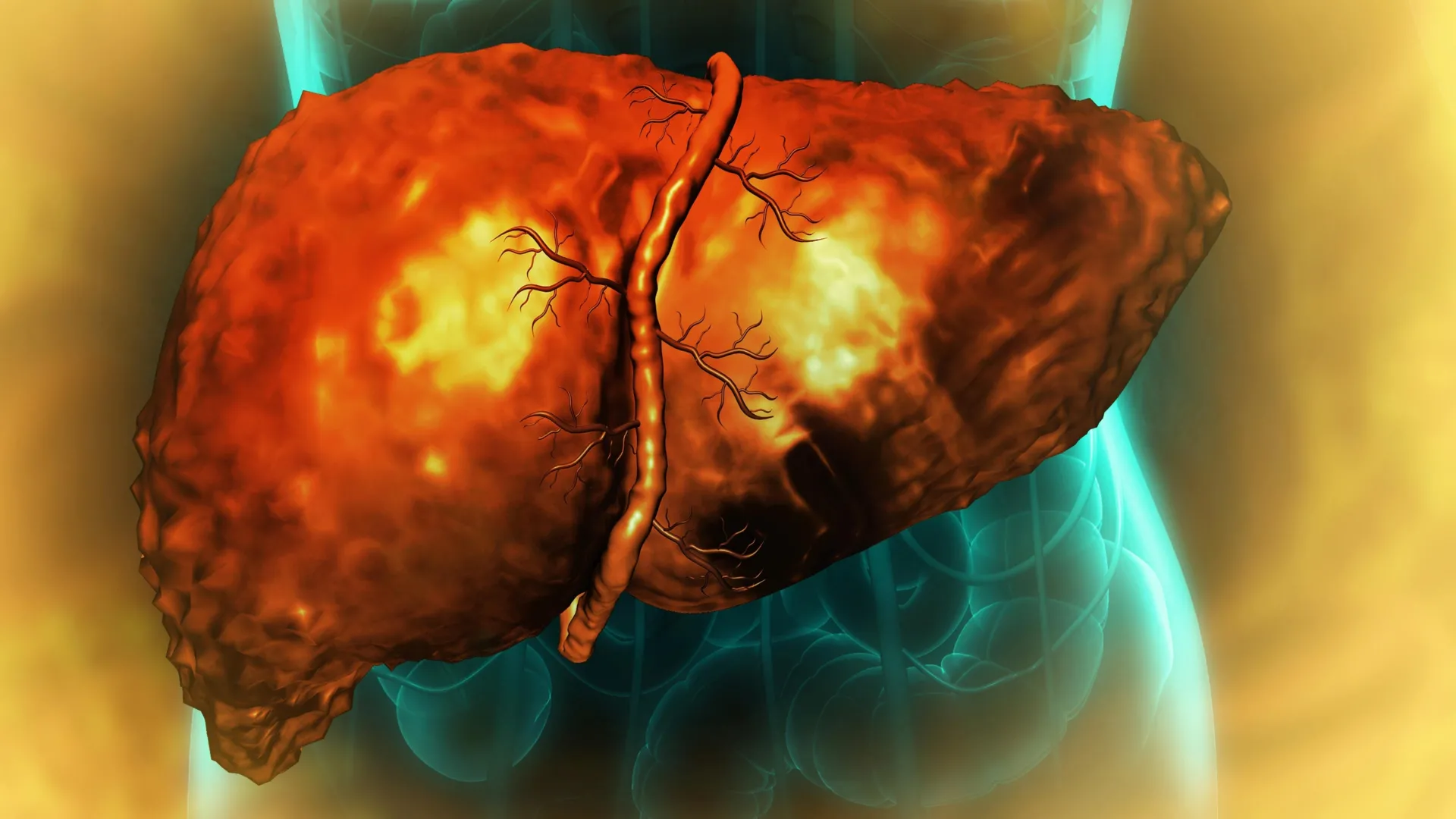
The liver, a remarkable organ celebrated for its regenerative prowess, can fall victim to a debilitating state of arrested development, even after an individual ceases alcohol consumption. New scientific inquiry elucidates a critical mechanism whereby alcohol-induced damage traps liver cells in an aberrant intermediate phase, rendering them incapable of fulfilling their normal functions or completing the vital process of tissue renewal. This cellular paralysis, previously an enigma, is now understood to be intricately linked to a cascade of inflammation that profoundly disrupts RNA splicing, a fundamental cellular operation essential for translating genetic blueprints into functional proteins.
This groundbreaking research, a collaborative effort involving scientists from the University of Illinois Urbana-Champaign, Duke University, and the Chan Zuckerberg Biohub Chicago, offers a paradigm shift in our understanding of alcohol-associated liver disease. The findings, meticulously detailed in the journal Nature Communications, illuminate not only the molecular underpinnings of this failure to heal but also pave the way for novel diagnostic markers and therapeutic interventions targeting severe liver ailments.
The liver’s extraordinary capacity to repair itself following significant injury, or even after surgical resection, sets it apart among major human organs. Under typical circumstances, surviving hepatocytes, the primary functional cells of the liver, can temporarily alter their specialized identities, proliferate, and subsequently mature to reconstitute lost tissue. However, this inherent resilience is critically compromised in the context of alcohol-associated liver disease, a condition that stands as the leading global cause of liver-related mortality, contributing to approximately three million deaths annually.
"We observed a clear impairment in liver function and regeneration in patients suffering from alcoholic hepatitis and cirrhosis, even when alcohol intake had been discontinued. The fundamental question of ‘why’ this occurred remained unanswered," explained Auinash Kalsotra, a professor of biochemistry at the University of Illinois and a co-leader of the study, alongside Anna Mae Diehl, a professor at Duke University School of Medicine. "At the advanced stages of these diseases, liver transplantation represents the sole life-saving intervention. Our pursuit was to unravel the underlying causes of liver failure, hoping to identify points of therapeutic intervention."
Professors Kalsotra and Diehl have dedicated years to deciphering the intricate molecular processes that govern hepatic regeneration. Their prior investigations established that during the regenerative phase, liver cells undergo a temporary reprogramming of their gene expression profiles. To initiate repair, mature hepatocytes revert to a state resembling fetal progenitor cells, which are less specialized and possess the capacity for division and new tissue generation. Following multiplication, these cells are meant to reverse this dedifferentiation process, ultimately regaining their mature, fully functional adult characteristics. This prior discovery served as the impetus for the researchers to investigate the specific molecular disruptions that derail this regenerative cycle in alcohol-induced liver pathology.
A striking observation emerged when comparing healthy liver tissue with samples obtained from individuals diagnosed with alcoholic hepatitis or cirrhosis. These diseased samples were acquired through an initiative supported by the National Institute on Alcohol Abuse and Alcoholism, a branch of the National Institutes of Health, at Johns Hopkins University Hospital. The analysis revealed a consistent anomaly: in the compromised livers, cells had initiated a transition away from their mature state towards a regenerative phenotype but were inexplicably unable to complete this transformation. Consequently, they became arrested in an intermediate, or "limbo," state.
"These cells were neither fully functional adult hepatocytes nor actively proliferating progenitor cells," elaborated Ullas Chembazhi and Sushant Bangru, graduate students at the University of Illinois and co-first authors of the study. "Their inability to perform their designated functions placed an increased burden on the remaining healthy cells, prompting them to attempt regeneration. However, this also led them to enter the same unproductive, quasi-progenitor state, ultimately precipitating liver failure." This creates a vicious cycle: as more cells succumb to this non-functional state, the burden on the dwindling population of healthy cells intensifies, increasing their risk of undergoing the same detrimental transformation.
To pinpoint the molecular culprits behind this stalled regeneration, the research team meticulously examined the proteins synthesized within liver cells and the messenger RNA (mRNA) molecules that convey genetic instructions from DNA to the protein-building machinery. RNA serves as a critical intermediary in this process. Before many mRNA molecules can be utilized, specific segments are excised and reattached in a process known as RNA splicing. The precise arrangement of these RNA segments is paramount, as it dictates the type of protein produced and its ultimate destination within the cell.
Rather than relying on conventional methods that quantify total RNA and protein levels, Professor Kalsotra’s team employed advanced deep RNA sequencing and sophisticated computational analyses to investigate the intricacies of RNA splicing. "Our comparative analysis revealed widespread RNA missplicing across thousands of genes in alcohol-related liver disease, significantly impacting the functional capabilities of numerous proteins," stated Professor Kalsotra, who also holds an affiliation with the Carl R. Woese Institute for Genomic Biology at Illinois. The magnitude of this misregulation was substantial, suggesting a broad disruption of protein function within damaged liver cells.
A key factor implicated in these widespread splicing errors was identified as a deficiency in a protein known as ESRP2. ESRP2 plays a crucial role in binding to RNA and ensuring its accurate splicing. The researchers observed significantly reduced levels of ESRP2 in alcohol-damaged liver cells. The ramifications of this deficiency extended beyond mere protein production; in numerous instances, the altered RNA sequences dictated incorrect localization of proteins within the cell.
"Proteins must function at specific cellular locations, a process governed by sequences within the protein itself that direct it to its designated site. We discovered that, frequently, the sequences responsible for protein localization were misspliced," explained Professor Kalsotra, who is also a member of the Chan Zuckerberg Biohub Chicago. "This underscores the importance of our multi-faceted analytical approach. While the quantity of RNA and protein might appear normal, their mislocalization due to splicing defects rendered them incapable of performing their intended roles. Crucially, key proteins essential for effective liver regeneration were found sequestered in the cytoplasm instead of migrating to the nucleus, where their function is required." The nucleus houses the cell’s DNA and is central to regulating gene activity, while the cytoplasm is the fluid-filled space where numerous other cellular processes occur. The mislocalization of vital proteins to the cytoplasm prevents them from executing their regenerative duties, even if present in adequate amounts.
To rigorously validate the role of ESRP2 deficiency in regeneration failure, the researchers conducted experiments with mice genetically engineered to lack the ESRP2-producing gene. These animal models exhibited patterns of liver injury and impaired regeneration that closely mirrored the observations in human patients with advanced alcoholic hepatitis. This finding prompted a crucial subsequent question: what leads to the reduction in ESRP2 levels in the first place?
The investigation traced the root cause of ESRP2 depletion back to chronic inflammation. The metabolic processing of alcohol by the liver triggers tissue damage, attracting immune cells and resident hepatic support cells to the injured sites. According to the study, these recruited cells release a surge of inflammatory and growth factors. The researchers demonstrated that these signaling molecules actively suppress both the synthesis and the functional activity of ESRP2.
The research team then explored the potential of interrupting these inflammatory signals to reverse the detrimental effects. In controlled laboratory settings using cultured liver cells, the scientists introduced a molecule designed to block the receptor for a key pro-inflammatory factor. Following this intervention, ESRP2 levels were restored, and RNA splicing patterns returned to a more normal state. This pivotal result suggests that the inflammatory pathway represents a promising therapeutic target. Instead of directly attempting to repair damaged liver tissue, future treatments could focus on disrupting the inflammatory signals that impede the cells’ innate regenerative capabilities. Furthermore, the researchers envision diagnostic applications, where abnormally spliced RNA molecules could serve as biomarkers to identify or monitor the progression of alcohol-associated liver disease.
"I am optimistic that these findings will serve as a catalyst for future clinical investigations," stated Professor Kalsotra. "We can leverage these misspliced RNAs as diagnostic indicators or develop therapeutic strategies to mitigate inflammation. By correcting these splicing defects, we may be able to enhance recovery and restore the function of damaged livers."
The extensive research team included biochemistry graduate students Diptatanu Das and Subhashis Natua from the University of Illinois; undergraduate students Katelyn Toohill, Ishita Purwar, and Anuprova Bhowmik from the University of Illinois; Brandon Peiffer and Zhaoli Sun from Johns Hopkins University School of Medicine; Aurelia Leona and Yogesh Goyal from Northwestern University; and Rajesh Dutta from Duke University School of Medicine. This significant research endeavor was made possible through the generous support of the National Institutes of Health, the Chan-Zuckerberg Biohub Chicago, the Duke Endowment, and the Muscular Dystrophy Association, with specific funding provided by various National Institutes of Health grants including R01-AA010154, R01-HL126845, R21-HD104039, R01-AA010154, 5R01-DK077794, 1R56-DK1343340, and R24 AA025017.
About the Author