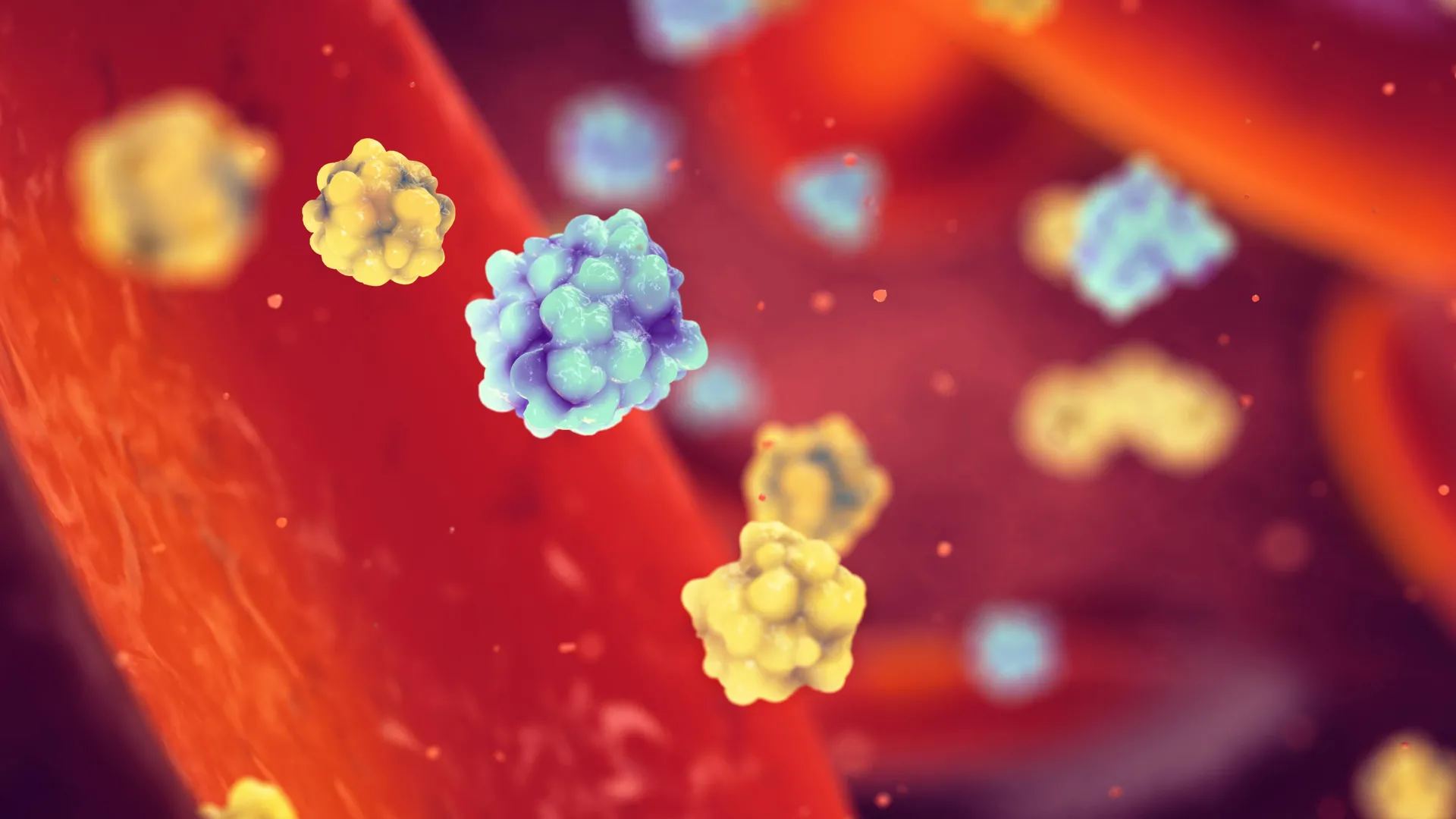
The intricate biological processes that govern cellular function often hinge on the precise three-dimensional conformation of proteins, akin to how a carefully folded piece of paper can transform into an elaborate origami creation. Within the specialized cells responsible for synthesizing insulin, this critical protein folding mechanism can falter, particularly as the body transitions through prediabetic states toward full-blown diabetes. When proteins, especially the precursor to insulin known as proinsulin, fail to achieve their correct molecular architecture, they accumulate in a misfolded state. This buildup exerts significant cellular stress, ultimately contributing to the dysfunction and eventual damage of pancreatic beta cells, the vital factories of insulin.
Recent investigations, spearheaded by a collaborative effort between researchers at Sanford Burnham Prebys Medical Discovery Institute and the University of Michigan, have illuminated the complex interplay of cellular components that orchestrate the delicate art of protein folding within these crucial insulin-producing cells. Published on June 1, 2026, in the esteemed journal Proceedings of the National Academy of Sciences, their findings offer a deeper understanding of how these cells maintain protein integrity and the cascade of events that ensue when this sophisticated system is disrupted. The implications of this research are profound, suggesting that bolstering the cell’s intrinsic protein-folding machinery could represent a novel strategy to safeguard these indispensable cells from the ravages of diabetes.
The pancreatic beta cells occupy a central role in glucose homeostasis, acting as sophisticated sensors that continuously monitor blood glucose levels. Upon detecting an elevation in blood sugar, these cells are prompted to ramp up their production of insulin, a hormone essential for facilitating the uptake of glucose by tissues and thereby restoring blood glucose to a healthy equilibrium. However, in the context of advancing diabetes, the relentless demand for insulin places an unsustainable burden on these beta cells, pushing them to their functional limits.
Previous scientific endeavors had already identified the aberrant folding of proinsulin as a significant contributor to the decline in beta cell function. It was understood that improperly shaped proinsulin molecules aggregate within these cells, creating a toxic environment that compromises their well-being. Yet, the precise molecular partners and their coordinated actions in managing this proinsulin misfolding crisis remained largely enigmatic.
"Our prior knowledge indicated that the cellular apparatus designed to prevent proinsulin misfolding relied on a key molecular chaperone, identified as binding immunoglobulin protein (BiP), in conjunction with a suite of auxiliary proteins known as cochaperones," explained Randal J. Kaufman, PhD, a distinguished professor within the Center for Metabolic and Liver Diseases at Sanford Burnham Prebys and the senior author of the study. "Our primary objective was to unravel the intricate mechanisms by which these collaborating proteins collectively ensure the correct folding of proinsulin and efficiently clear any misfolded anomalies. These processes are absolutely fundamental to the sustained health and operational capacity of insulin-producing cells."
To meticulously trace the interactions of BiP within beta cells, the research team employed a sophisticated genetic engineering approach in mice. They modified the beta cells of these animals to express a version of BiP that carried an appended molecular tag. This tag, consisting of three tandem copies of an eight-amino-acid sequence known as a 3xFLAG-tag, functioned as a highly visible molecular beacon. This innovation significantly simplified the researchers’ ability to detect, isolate, and study BiP during their experimental analyses.
The experimental results strongly implicated a particular cochaperone protein, designated as p58IPK, as playing a disproportionately crucial role in this cellular drama. When the researchers experimentally deactivated p58IPK in two distinct beta cell lines, they observed a marked increase in the accumulation of misfolded proinsulin. This observation was further corroborated by studies conducted on mice that were genetically engineered to be incapable of producing p58IPK. These animals exhibited a significant reduction in the synthesis of both proinsulin and mature insulin within their beta cells, providing compelling evidence for p58IPK‘s essential function.
The scientific team then proceeded to reintroduce functional p58IPK into one of the genetically modified cell lines where it had been absent. This restoration of the cochaperone dramatically improved the cells’ capacity to accurately fold proinsulin and efficiently transport it through the cellular pathways, concurrently diminishing the problematic aggregation of improperly folded proinsulin molecules.
However, the study also revealed that p58IPK, while critical, could not unilaterally substitute for the indispensable role of BiP. The observed improvements in proinsulin processing were contingent upon the presence of BiP; without it, the beneficial effects of p58IPK were not realized. Further investigations sought to ascertain whether augmenting the levels of BiP could compensate for the absence of p58IPK. The findings indicated that cells engineered to produce elevated quantities of BiP but lacking p58IPK experienced only marginal enhancements in proinsulin folding and its subsequent export from the cell. The most substantial improvements were documented when both BiP and p58IPK were present at their normal physiological concentrations.
"Our findings underscore the principle that, much like a lone tennis player attempting to navigate a doubles match, BiP cannot independently maintain the correct structural integrity of proinsulin," remarked Insook Jang, PhD, a senior scientist in the Kaufman laboratory and the lead author of the published research. "This highlights a critical interdependence between these molecular players."
Beyond BiP and p58IPK, the investigators also identified additional protein partners involved in the multifaceted process of proinsulin folding, its subsequent trafficking, and the cellular mechanisms responsible for detecting and managing misfolded proteins. The precise influence of these newly identified proteins on insulin production and the trajectory of diabetes progression warrants further in-depth research.
"Our studies collectively emphasize that the process of proinsulin folding is susceptible to many of the same cellular stressors that precipitate beta cell failure in the context of type 2 diabetes," Professor Kaufman reiterated, underscoring the broad relevance of their findings.
The therapeutic landscape for diabetes currently comprises treatments that primarily focus on managing blood glucose levels through mechanisms such as enhancing glucose uptake by peripheral tissues or stimulating the pancreas to release more insulin. Crucially, these existing pharmacological interventions do not directly address the underlying protein-folding defects that are increasingly recognized as a significant contributor to beta cell demise. To date, no therapies have been specifically designed to rectify proinsulin misfolding with the aim of preserving the health and functionality of insulin-producing beta cells.
"Should we succeed in deciphering how to modulate the synchronized activity of BiP, a central orchestrator of proinsulin folding, we may uncover a highly promising therapeutic strategy," stated Professor Kaufman. "Such an approach could offer an opportunity for early intervention, potentially preventing or significantly mitigating the damage inflicted upon insulin-producing cells, thereby offering a new paradigm in diabetes management."
Additional contributors to this significant study include Alec Duffey and Pamela Itkin-Ansari from Sanford Burnham Prebys, and Peter Arvan from the University of Michigan. The research was generously supported by funding from the National Institutes of Health, specifically grants from the National Institute of Diabetes and Digestive and Kidney Diseases, the National Cancer Institute, and Breakthrough T1D (formerly known as the Juvenile Diabetes Research Foundation).
About the Author