Injectable Biomaterial Facilitates Neural Tissue Regeneration and Functional Recovery Post-Ischemic Stroke
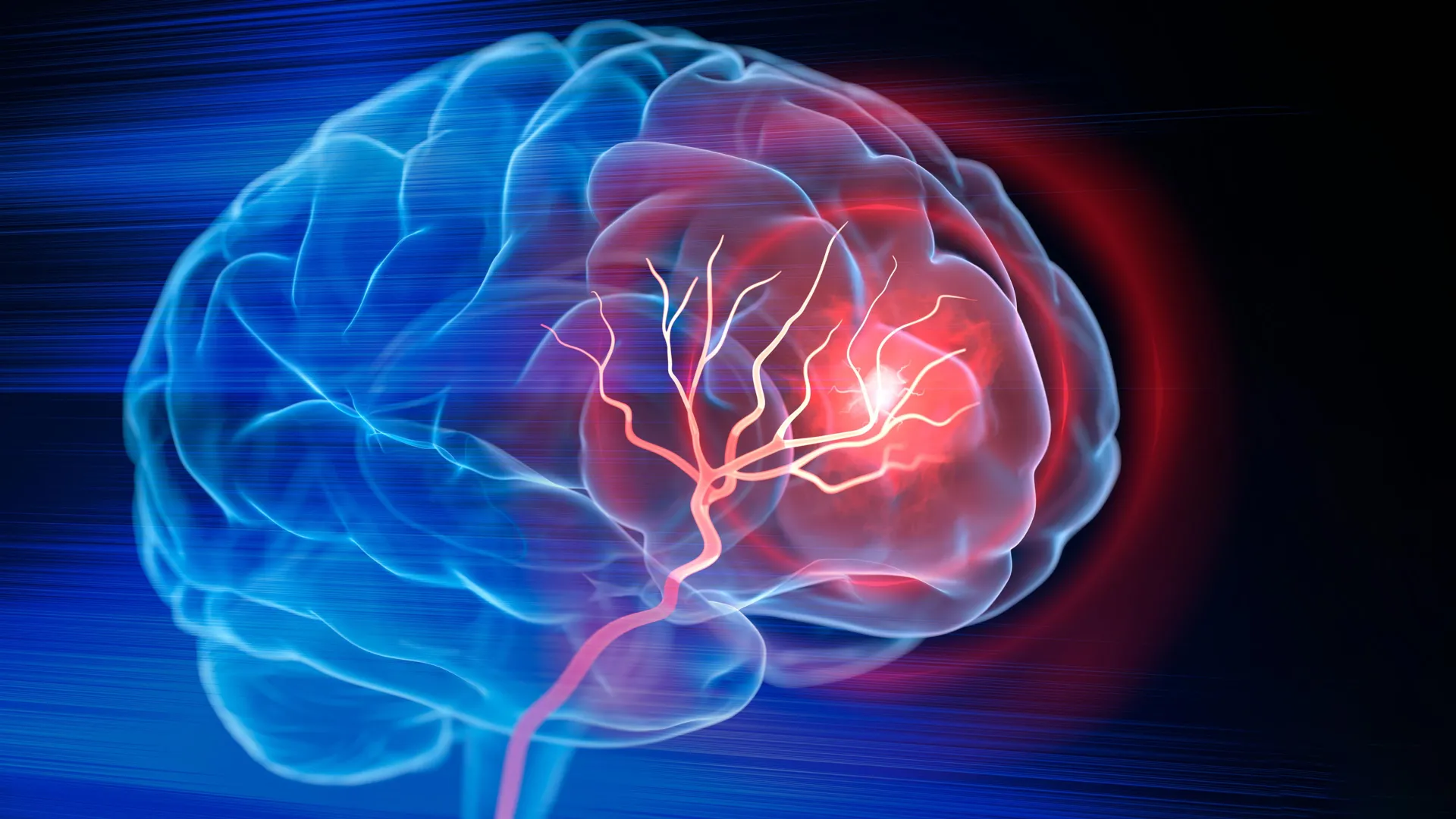
Biomedical engineers at Duke University have engineered an innovative injectable biomaterial designed to foster neural tissue regeneration and facilitate functional recovery following ischemic stroke events. In preclinical investigations utilizing rodent models, this sophisticated material demonstrated a remarkable capacity to transform the void left by destroyed brain tissue into a milieu conducive to intricate repair processes. This advanced therapeutic approach not only stimulated the body’s endogenous immune response but also actively promoted the development of new vasculature, supported critical alterations within the neural architecture, and ultimately led to significant improvements in motor function among the test subjects. The comprehensive findings detailing this breakthrough have been formally presented and published in the esteemed scientific journal Cell Biomaterials.
The profound challenge of addressing the neurological deficits left in the wake of ischemic strokes has long been a central focus in neuroscientific research and clinical practice. Each year, millions of individuals worldwide experience these debilitating events, which are characterized by the sudden interruption of blood flow to a portion of the brain, typically due to a blood clot. While immediate interventions, such as the administration of clot-dissolving pharmaceuticals or mechanical thrombectomy procedures to physically extract the obstruction, can be instrumental in salvaging brain tissue that remains viable, their efficacy wanes once neuronal death has irrevocably occurred. The permanent loss of brain tissue, particularly in severe stroke cases, can result in the formation of significant cavities within the brain parenchyma. Following the restoration of circulation and the removal of the occluding clot, the trajectory of recovery predominantly relies on intensive rehabilitation therapies. Although rehabilitation plays a crucial role in optimizing the adaptability of existing neural circuits and compensating for lost function, it does not directly address the fundamental task of rebuilding the compromised anatomical region.
"Once brain tissue has been lost, simply restoring blood flow is no longer a sufficient intervention," explained Dr. Tatiana Segura, the Robert Plonsey Distinguished Professor of Biomedical Engineering at Duke University and a leading figure in this research. "Our overarching objective is to engineer the injured cerebral space in such a manner that it actively orchestrates the collaborative efforts of immune, vascular, and neural repair mechanisms."
The research team embarked on a mission to construct an internal environment within the stroke-induced cavity that could simultaneously support a multitude of repair modalities. Their innovative solution involved the development of microporous annealed particle (MAP) scaffolds. These scaffolds are fabricated from an assembly of individual hydrogel microparticles, meticulously engineered to form a porous, three-dimensional framework. This open, interconnected architecture provides an essential structural substrate, offering resident and infiltrating cells a physical scaffold upon which they can migrate, proliferate, and actively engage in the rebuilding of neural tissue. Building upon prior successful applications of this biomaterial, the researchers sought to investigate whether the body’s inherent immune system could be harnessed to both guide and amplify the regenerative process.
A pivotal component of their strategy involved the strategic utilization of astrocytes, a class of glial cells characterized by their star-like morphology. Astrocytes are known to perform a multitude of critical functions in the healthy brain and exhibit a pronounced reactive response following neurological injury. A primary mechanism through which astrocytes communicate with neighboring cells involves the release of extracellular vesicles (EVs). These minuscule lipid-bound packages are instrumental in intercellular communication, carrying a diverse cargo of proteins, lipids, and genetic material that can profoundly influence the behavior and function of recipient cells.
The researchers meticulously collected EVs from astrocytes cultured in laboratory settings and subjected them to rigorous testing with a panel of signaling molecules specifically designed to attract immune cells, promote the formation of new blood vessels, and enhance overall functional recovery. Rather than indiscriminately introducing these EVs into the compromised brain region, the scientists ingeniously devised a method to chemically conjugate them to the surface of the hydrogel microparticles constituting the MAP scaffold. This innovative approach ensured that the therapeutic signaling molecules remained localized and concentrated within the scaffold’s intricate network, thereby significantly increasing the probability of their interaction with incoming cellular components and optimizing their biological impact.
"We are not merely introducing an inert material into the brain," Dr. Segura emphasized. "Instead, we are actively engineering a highly localized environment that possesses the capacity to coordinate multiple facets of the repair response." Among the various signaling combinations investigated, a particular pairing of Interleukin-4 (IL-4) and C1q emerged as exceptionally effective in attracting potent immune cells, including macrophages and, surprisingly, a remarkably persistent population of neutrophils, into the injured cerebral area.
The emergent role of neutrophils in this regenerative process presented an unexpected but significant finding. While neutrophils are conventionally associated with inflammatory responses and tissue damage during the acute phase of a stroke, the new findings suggest a more nuanced and potentially beneficial contribution at later stages of recovery. The study indicated that, when presented with the appropriate signaling cues and the supportive microenvironment provided by the engineered scaffold, neutrophils could actively contribute to tissue repair rather than exacerbating damage. To rigorously assess the importance of these cells, the researchers deliberately reduced the population of neutrophil-rich immune cells in their experimental models. This manipulation led to a substantial decline in the rate of new blood vessel formation and a notable reduction in the remodeling of the scaffold itself, underscoring the critical role of these immune cells in orchestrating the healing cascade.
"This particular result fundamentally alters our understanding of the role neutrophils play in the aftermath of a stroke," stated Dr. Shangjing Xin, the lead scientist on this study and a postdoctoral fellow within Dr. Segura’s laboratory. "Their function appears to be highly context-dependent, influenced by their timing of arrival, their precise location within the injured tissue, and the specific signals they receive from their immediate surroundings. Our research demonstrates a potential engineering strategy to effectively recruit and retain these cells at the opportune moments for maximal therapeutic benefit."
Concurrently with the influx of immune cells, a robust formation of new blood vessels was observed throughout the entirety of the stroke cavity. Further histological analysis revealed a significant increase in axonal fibers, the vital conduits responsible for transmitting neural signals, both within the confines of the injured region and extending into the surrounding brain tissue. These profound biological transformations were directly correlated with marked improvements in the animals’ motor capabilities. In a standard grid-walking test, designed to quantify errors in forelimb placement, mice treated with the optimized scaffold exhibited significantly enhanced performance. By the eight-week mark of the study, their performance on this task was statistically indistinguishable from that of healthy control subjects, and this functional recovery proved to be enduring for the remainder of the experimental period.
Crucially, the research team also conducted experiments to ascertain whether the extracellular vesicles could elicit similar regenerative effects in the absence of the biomaterial scaffold. These comparative trials revealed that EVs administered alone, without the presence of the MAP scaffold, failed to induce comparable levels of vascular repair or neural regeneration. This critical observation strongly suggests that the biomaterial’s contribution extended far beyond its role as a simple delivery vehicle for therapeutic signals. The synergistic interplay between the scaffold’s inherent porous architecture, which facilitated cellular infiltration and interaction, and its capacity to maintain a concentrated milieu of EV-derived signaling molecules within the damaged zone, appeared to be indispensable for initiating and sustaining the observed repair processes.
Despite the highly promising nature of these preclinical findings, it is essential to emphasize that this therapeutic approach remains in its nascent stages of development. To date, the efficacy of this treatment has been rigorously evaluated solely in mouse models, involving the direct injection of the biomaterial into the precisely identified lesion site within the brain. Extensive further research will be imperative to comprehensively assess the safety profile of this intervention, to elucidate with greater precision the intricate mechanisms by which diverse immune cell populations contribute to the recovery process, and to definitively determine whether this promising treatment translates effectively to larger animal models that more closely recapitulate the complexities of human stroke.
Currently, the research team obtains the necessary EVs from primary rat astrocytes. As a critical next step in advancing the clinical translatability of this technology, Dr. Segura’s laboratory is actively investigating the use of EVs derived from human induced pluripotent stem cell-derived astrocytes. Such cells hold the potential to provide a more scalable and clinically relevant source of EVs, while simultaneously affording researchers greater control over the specific signaling molecules encapsulated within these vesicles.
"The restoration of a complex biological ecosystem, such as the brain, cannot be achieved simply by containing the initial damage," Dr. Segura articulated. "It necessitates the deliberate creation of conditions that foster the re-emergence and proliferation of life. This is precisely how we conceptualize the transformation of the stroke cavity. The material itself is not intended to replicate the intricate structure of the brain, but rather to cultivate an environment wherein the body’s own cellular components can ingress, engage in sophisticated communication, and actively participate in the reconstruction of vascularized, functional tissue."

Related News