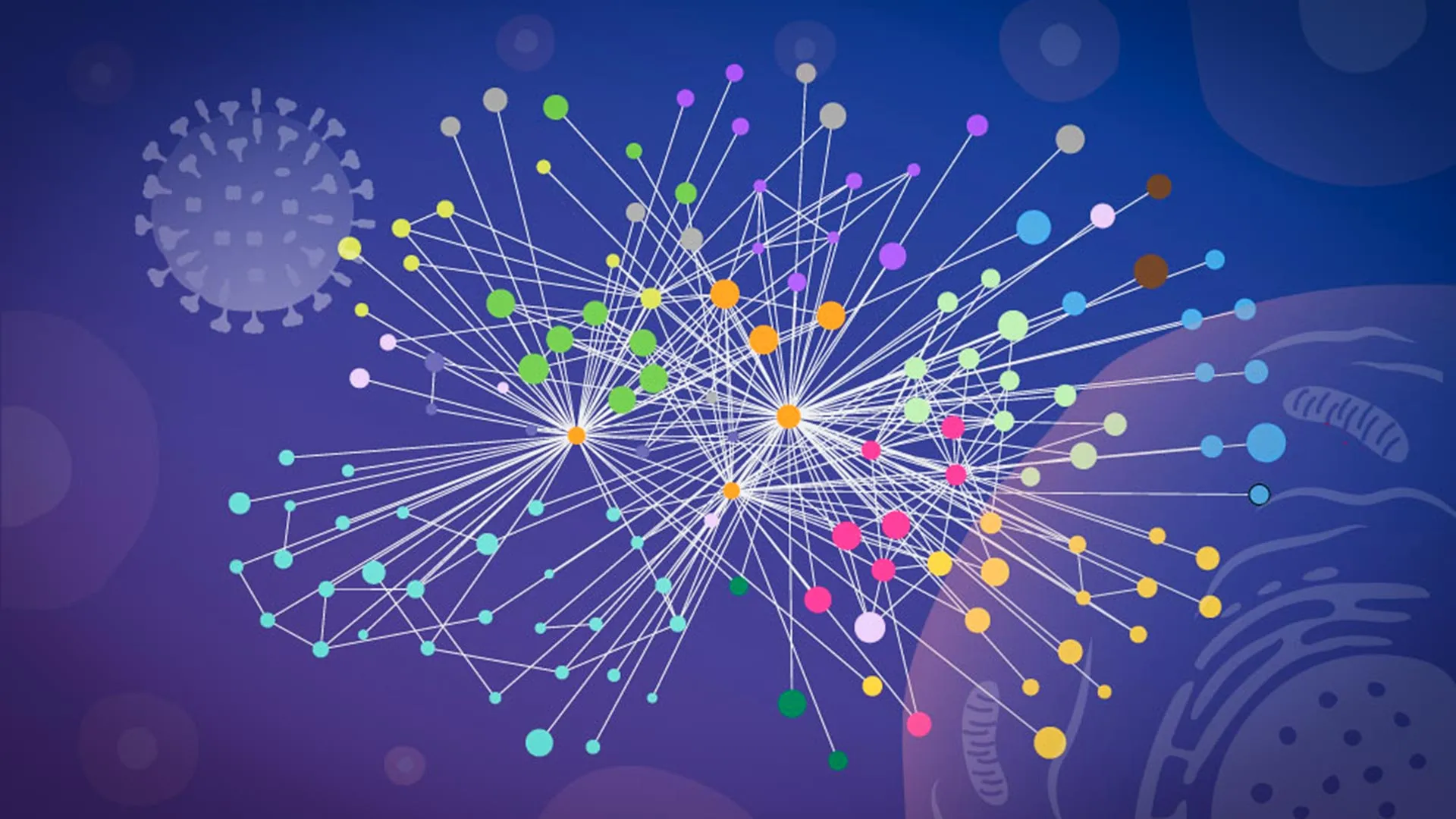
Influenza A, a perennial global health concern responsible for millions of severe illnesses and hundreds of thousands of fatalities annually, and a historical architect of devastating pandemics, including the infamous 1918 Spanish Flu, has now been meticulously mapped at a molecular level within living human cells. This groundbreaking achievement, spearheaded by a collaborative effort between researchers at EMBL Hamburg and the Leibniz Research Institute for Molecular Pharmacology (FMP), provides an unprecedentedly detailed visualization of how the virus orchestrates the hijacking of its host’s cellular machinery. Unlike previous investigations that often dissected cells, compromising the integrity of delicate molecular interactions, this new study employed a sophisticated workflow allowing for direct observation of protein engagements within intact, infected cellular environments.
The fundamental modus operandi of influenza A involves the virus’s entry into a host cell, followed by the release of its genetic material – RNA – which carries the blueprint for synthesizing a select group of viral proteins. These viral components then infiltrate the host cell, subtly rerouting its intricate molecular systems to transform the cell into a clandestine factory churning out new viral progeny. Understanding the precise nature of this cellular invasion, specifically which viral proteins engage with human proteins, the exact locations of these interactions, and how the virus leverages these connections to facilitate its replication, is paramount for developing more potent influenza vaccines and effective antiviral therapies.
This pioneering research represents the first large-scale mapping of direct physical contacts between influenza viral proteins and human cellular proteins within intact, infected cells. The level of structural resolution achieved is sufficiently precise to enable scientists to construct models illustrating the potential spatial arrangements of these interacting protein pairs. Jan Kosinski, a Group Leader at EMBL Hamburg and the Centre for Structural Systems Biology (CSSB), highlighted the significance of this novel approach, stating that it offers a new paradigm for studying flu-host interactions in their natural setting with remarkable structural clarity. He further elaborated that while the current findings represent a snapshot of a specific moment during the infection process, this work opens avenues for investigating the complete spectrum of flu-host interactions throughout the entire infection cycle.
The inherent difficulty in tracking protein-protein interactions during an active viral infection has been a significant hurdle in virology research. Historically, many studies relied on biochemical methodologies that necessitated breaking open cells to measure protein contacts. This destructive process, however, carries the inherent risk of distorting the true picture of what transpires within a living cell. Once cellular compartments are compromised, proteins that were spatially separated in their native environment might come into close proximity in the laboratory setting. Conversely, weak, transient, or location-specific interactions could be lost altogether, making it challenging for researchers to definitively ascertain which connections were genuinely present during the actual infection.
The critical breakthrough for this research team emerged when they discovered that their collaborators, Boris Bogdanow and Fan Liu, at FMP Berlin, had ingeniously adapted a long-established technique known as cross-linking mass spectrometry (XL-MS). This specialized version of XL-MS was meticulously tailored for studying virus-infected cells, enabling the capture of protein interactions occurring only briefly or within specific cellular regions. Bogdanow, now a Junior Research Group Leader at the Institute of Virology, Charité – Universitätsmedizin Berlin, explained that XL-MS allows for the direct capture of protein-protein interactions within intact, infected cells, simultaneously providing structural insights into the mechanics of these engagements. This capability offers a profound understanding of the interface between the virus and the human cell, potentially revealing actionable targets for future pharmaceutical interventions through structural modeling.
To further elucidate these interactions, the researchers integrated their XL-MS experimental data with advanced computational structural modeling techniques. This synergistic approach allowed them to not only identify the specific viral and human proteins that interact but also to estimate their relative positions when engaged. The structural models were constructed using a modified version of AlphaFold, the renowned protein structure prediction algorithm recognized with a Nobel Prize. Kosinski emphasized the pivotal advantage of this adapted AlphaFold methodology, explaining that it permitted the direct integration of their experimental cross-linking data into the structural modeling process. This crucial input guided the model by indicating which regions of the viral and host proteins were in close proximity within infected cells, proving particularly beneficial for analyzing virus-host complexes, which are often challenging to predict with high accuracy.
The findings, published in the prestigious journal Nature Microbiology, unveiled two significant strategies employed by influenza A in its successful takeover of host cells. The first strategy centers on hemagglutinin, a protein adorning the outer surface of the influenza virus, which facilitates its attachment to and entry into host cells. The researchers tracked hemagglutinin’s journey through the cell’s intricate internal transport and processing network – a complex system of compartments responsible for folding, modifying, and directing proteins to their designated locations. Their analysis revealed that several human proteins played a crucial role in the correct folding and modification of hemagglutinin during the infection process, including some host proteins whose functions were previously not well understood.
The second striking discovery pertained to paraspeckles, small, droplet-like structures residing within the cell’s nucleus. The research team observed that influenza A infection induced the dissolution of these paraspeckles. Upon the breakdown of paraspeckles, RNA-binding proteins, which were previously sequestered within them, were released. The virus can then potentially utilize these liberated proteins to bolster its own replication efforts. Iuliia Kotova, a former predoctoral fellow in the Kosinski Group at EMBL Hamburg and the first author of the publication, expressed her astonishment at the paraspeckle findings, noting that their consistent dissolution across various cell lines and influenza strains suggested it was not merely a byproduct of infection but likely a deliberate viral strategy.
The disruption of paraspeckles may offer the influenza virus multiple advantages. Kosinski speculated that a secondary benefit for the virus could stem from paraspeckles’ known role in cellular stress responses and antiviral gene regulation. By dismantling these structures, the virus could potentially weaken the cell’s intrinsic defense mechanisms.
This complex undertaking was made possible through the synergistic sharing of advanced technologies and specialized expertise across three distinct institutions. The crucial cross-linking mass spectrometry experiments were conducted at Charité in Berlin, while comprehensive glycoproteomics analyses were performed at the EMBL Proteomics Core Facility. The computational structural modeling was carried out on the EMBL Compute Cluster, and advanced microscopy imaging was conducted at the Advanced Light and Fluorescence Microscopy (ALFM) Facility at CSSB.
This study underscores the profound insights gained by examining molecular contacts within intact infected cells, illuminating both the location and the mechanisms by which viruses seize control of human cellular machinery. The researchers posit that this "mapping in context" approach holds significant promise for unraveling the operational strategies of other viruses as well. Kosinski remarked that while the specific host factors and mechanisms may vary between different viruses, their overarching methodology – a combination of in-cell cross-linking, structural modeling, and targeted cell-biology investigations to map native virus-host interactions at specific stages of infection – is broadly applicable.
Although this particular study utilized a laboratory-adapted strain of influenza, the researchers are confident that the same methodology can be extended to investigate viruses with greater pandemic potential. Bogdanow concurred, stating that this research lays the essential groundwork for applying their methodology to viruses of significant pandemic concern, such as H5N1, and for uncovering the intricate interaction networks that facilitate their proliferation within human cells.
About the Author