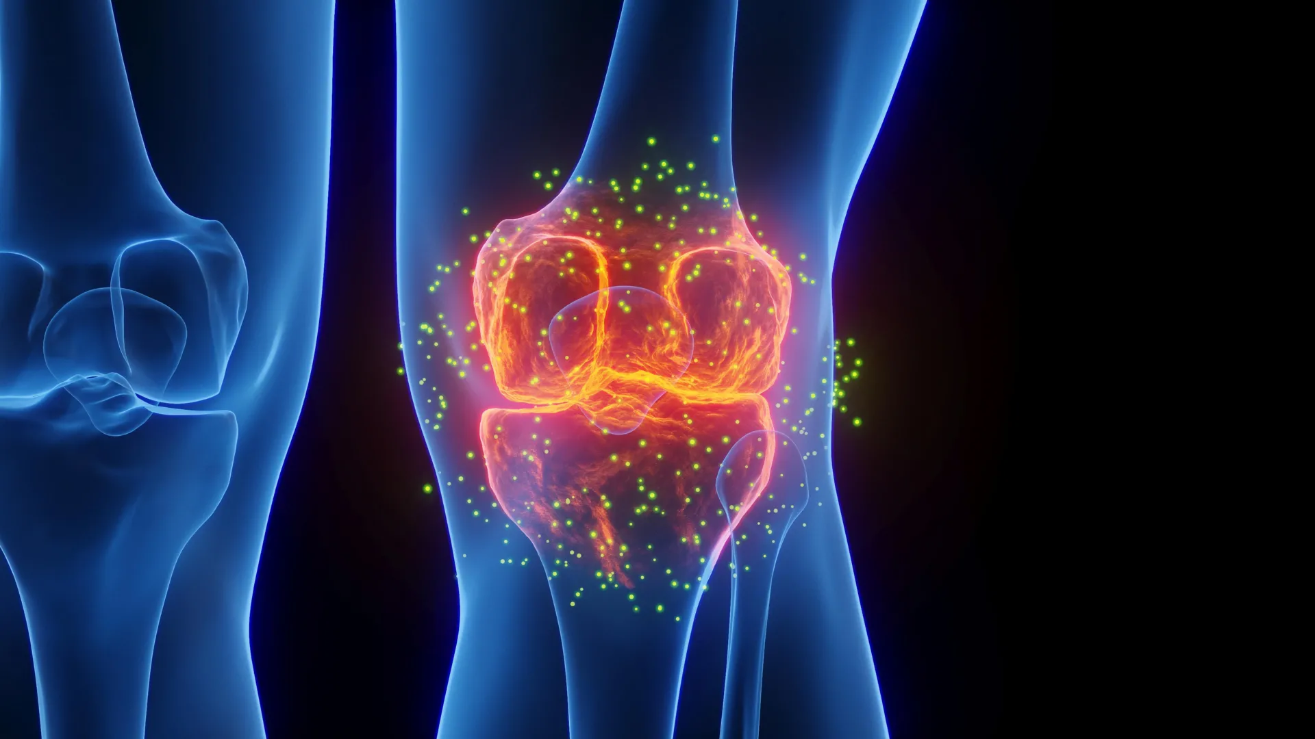
Post-traumatic osteoarthritis (PTOA), a debilitating and chronic condition affecting millions globally, frequently emerges following acute joint injuries such as ligament tears, meniscal damage, or fractures. Unlike its age-related counterpart, PTOA often strikes younger, active individuals, leading to a progressive deterioration of joint cartilage, persistent pain, reduced mobility, and a significant decline in quality of life. Current therapeutic strategies primarily focus on managing symptoms or surgically repairing immediate damage, with limited options available to effectively prevent or halt the underlying degenerative process. This challenging clinical landscape underscores an urgent need for innovative interventions that can intervene early and alter the disease trajectory. In a significant stride towards addressing this gap, a multidisciplinary research team at The University of Alabama in Huntsville (UAH), a constituent institution of The University of Alabama System, has unveiled compelling findings suggesting that continuous low-intensity ultrasound could represent a novel, non-pharmacological approach to prevent the onset and progression of PTOA by actively reshaping the body’s immune response.
The groundbreaking research, recently detailed in the esteemed scientific journal Scientific Reports, a Nature publication, proposes that this non-invasive acoustic technology possesses the remarkable capability to steer the body’s immune system away from a prolonged inflammatory state—a key driver of joint degradation—and towards a more reparative and regenerative healing pathway. This potential shift offers a beacon of hope for improving long-term outcomes for individuals sustaining joint trauma, potentially mitigating the development of chronic joint disease.
At the core of this investigation lies the intricate role of macrophages, specialized immune cells that are indispensable players in both the inflammatory response to injury and the subsequent processes of tissue repair. These versatile cells exhibit a remarkable plasticity, capable of adopting distinct functional phenotypes depending on the microenvironment and signals they receive. Dr. Anuradha Subramanian, a distinguished professor of chemical and materials engineering at UAH and the principal investigator for this study, elaborated on their critical function. "Following any tissue injury, the body swiftly mobilizes a population of pro-inflammatory ‘defender’ macrophages, often categorized as M1-like, to the site of damage," Dr. Subramanian explained. "Their primary mission is to clear cellular debris, pathogens, and initiate the initial stages of inflammation necessary for healing. Concurrently, or in a subsequent phase, ‘healer’ macrophages, known as M2-like, arrive to facilitate tissue regeneration and resolution of inflammation." The delicate balance between these two macrophage phenotypes is crucial for effective healing. However, as Dr. Subramanian emphasized, "a persistent dominance of these M1-like defender macrophages can establish a chronic inflammatory milieu that actively contributes to tissue damage and, critically, to the development of conditions like post-traumatic osteoarthritis."
The central hypothesis guiding the UAH team’s work was whether targeted application of continuous low-intensity ultrasound could actively influence these immune cells, encouraging them to transition from an inflammatory (M1-like) state to one that vigorously promotes healing and tissue repair (M2-like). Dr. Satyaki Roy, a professor of mathematical sciences who contributed advanced computational and statistical analyses to the study, underscored the clinical relevance of this approach. "Chronic inflammation is unequivocally recognized as a primary catalyst in the pathogenesis of post-traumatic osteoarthritis," Dr. Roy stated. "The relentless inflammatory cycle restricts the body’s natural capacity for tissue regeneration and accelerates the degenerative processes within the joint. Our team’s keen interest in continuous low-intensity ultrasound stems from its promise as a non-pharmacological, entirely non-invasive intervention that could potentially regulate immune cell behavior and cultivate a more reparative healing environment within injured joints."
To investigate this complex biological interaction, the UAH researchers embarked on a meticulously designed study, integrating sophisticated biological experiments with cutting-edge computational analyses. The biological groundwork was meticulously laid by Dr. Shahid Khan during his doctoral research, while graduate student Owen Trippany also made significant contributions to the collaborative effort. A critical aspect of their methodology involved creating a laboratory model that more accurately replicated the biochemical environment of an injured joint, moving beyond conventional, generalized inflammatory triggers. Instead of relying solely on standard methods to induce inflammation, the team utilized fibronectin fragments—specific molecules naturally generated as damaged joint tissue breaks down following trauma. This innovative approach yielded a model that much more closely mirrors the actual biological cascades and cellular signaling that unfold in a real-world joint injury scenario, thereby enhancing the translational relevance of their findings.
Beyond the biological modeling, the team employed a powerful combination of transcriptomics and an advanced computational method known as differential clustering. Transcriptomics, the large-scale study of gene activity within cells, provides a snapshot of which genes are turned "on" or "off" and to what extent. However, rather than merely analyzing individual gene expression in isolation, the differential clustering technique allowed the researchers to identify groups of genes whose activity patterns changed in a coordinated fashion in response to the ultrasound treatment. "This sophisticated analytical approach was pivotal," Dr. Roy explained. "It enabled us to move beyond simply identifying individual genes that were altered and instead provided a holistic understanding of how entire networks and groups of genes collectively modified their coordinated behavior in response to continuous low-intensity ultrasound stimulation. This offers a far more comprehensive picture of the cellular response than traditional gene-by-gene analysis."
The results of these rigorous investigations proved highly encouraging. The application of continuous low-intensity ultrasound consistently led to a measurable reduction in the biological markers that are intrinsically linked to inflammation. Simultaneously, the researchers observed a significant increase in the markers associated with a more reparative, M2-like macrophage state. These findings strongly suggest that the acoustic stimulation effectively modulated the phenotypic switch in macrophages, directing them away from destructive inflammation and towards a constructive healing phase.
While these findings are presently confined to carefully controlled laboratory experiments, their implications are profound. They open a compelling new vista for non-drug, non-invasive technologies to potentially influence fundamental immune cell behavior and substantially improve the healing process following joint injuries. The UAH researchers envision that this technique could ultimately be integrated into future therapeutic protocols specifically designed to slow or even prevent the relentless progression of osteoarthritis and significantly enhance recovery trajectories after various forms of joint trauma. The potential to offer patients a non-pharmacological alternative or adjunct therapy for a condition that currently has limited preventative options is immense.
Looking ahead, the UAH team has already charted the course for the next critical phases of their research. "The immediate next steps involve validating these promising findings in relevant animal models of early post-traumatic osteoarthritis," Dr. Subramanian articulated. "This will be crucial for understanding the in-vivo effects of ultrasound-based immune modulation. Concurrently, we will be meticulously studying how this acoustic intervention affects long-term tissue repair and regeneration within the complex environment of injured joints." This planned progression from in vitro studies to animal models represents a vital step on the long but hopeful journey towards potential clinical translation. If successful, this innovative approach could revolutionize the management of joint injuries, offering a proactive strategy to mitigate the onset of debilitating osteoarthritis and dramatically improve the quality of life for countless individuals worldwide. The prospect of harnessing sound waves to orchestrate cellular healing underscores a truly exciting frontier in regenerative medicine and musculoskeletal health.
About the Author