
Chronic discomfort in the lower back and knees represents a prevalent challenge for a significant portion of the adult population, particularly those navigating midlife and beyond. While conventional approaches frequently target the sites of pain directly, a growing body of evidence, supported by clinical experience, points to an often-overlooked area as a primary driver of these pervasive issues: the hip joint. Rather than solely a powerful engine for movement, the hip functions as an intricate nexus of stability and force distribution, whose subtle dysfunctions can propagate adverse effects throughout the musculoskeletal system. Understanding and addressing the holistic mechanics of the hip is paramount for individuals seeking to alleviate persistent pain and sustain functional independence as they age.
The human hip is far more than a simple ball-and-socket joint; it operates as a sophisticated "centration joint." Its fundamental responsibility lies in maintaining the optimal alignment of the femoral head precisely within the acetabulum, the socket of the pelvis. This precise centering is crucial for efficient load transfer and seamless movement. The hip’s operational integrity relies on a complex "relational system" encompassing not just the strength of individual muscle groups, but also the balanced tension within the joint capsule, the supportive role of ligaments, the precise alignment of the femoral head, the intricate neurological feedback, and the integrated load-bearing capacity of the fascial network. When any constituent element of this multifaceted system deviates from its optimal state—whether through weakness, stiffness, or altered neural patterning—the body’s inherent design for distributing forces is compromised. This breakdown inevitably leads to the adoption of compensatory movement patterns, placing undue stress on adjacent structures, most notably the lumbar spine and the knees, which then begin to manifest symptoms of pain and instability. For individuals progressing through their later decades, a primary focus shifts from merely building gross muscular strength to meticulously restoring segmental hip function and ensuring robust fascial continuity. When the hip regains its innate capacity to absorb and efficiently transmit forces, the broader biomechanical system often experiences a remarkable reduction in overall stress and inflammation.
A foundational approach to restoring hip health and, by extension, mitigating lower back and knee pain, involves several interconnected strategies, each targeting a specific aspect of the hip’s complex function. These strategies move beyond superficial strengthening, delving into the nuanced interplay of muscles, fascia, and neural pathways.
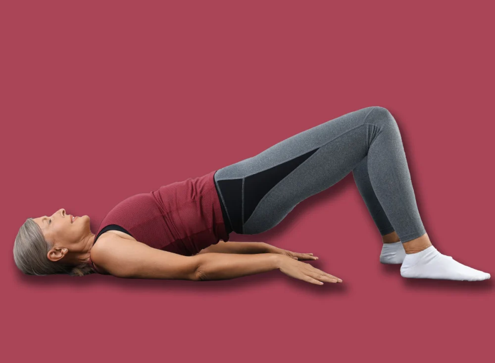
I. Re-establishing Gluteal Stabilizer Competence
One of the initial areas often identified as compromised in individuals experiencing referred pain is the functional integrity of the gluteal system’s stabilizers, specifically the gluteus medius and the deep lateral rotators. These muscles are not merely responsible for hip abduction or external rotation; their role extends critically to stabilizing the pelvis during locomotion and maintaining precise control over femoral rotation. From a comprehensive anatomical perspective, these muscles are integral components of the lateral fascial chains, which establish crucial connections between the hip, the thoracolumbar fascia of the lower back, and even extending to the contralateral shoulder.
When these vital lateral fibers experience a decline in their timely activation or overall recruitment capacity, a common consequence is an observable drop in the pelvis during the gait cycle. This phenomenon is clinically recognized as a Trendelenburg sign, indicating an imbalance that precipitates a lateral shift in the pelvis’s movement. Such a shift generates rotational stress that cascades down into the knee joint, simultaneously imposing significant shear forces on the lumbar spine. Over time, these repetitive, misaligned stresses contribute directly to the development of chronic pain and degenerative changes in these vulnerable areas. Targeted activation and strengthening of these gluteal stabilizers are therefore crucial, not just for hip movement, but for establishing a stable base that prevents detrimental force transference to the lower back and knees, thereby enhancing walking efficiency and reducing injury risk.
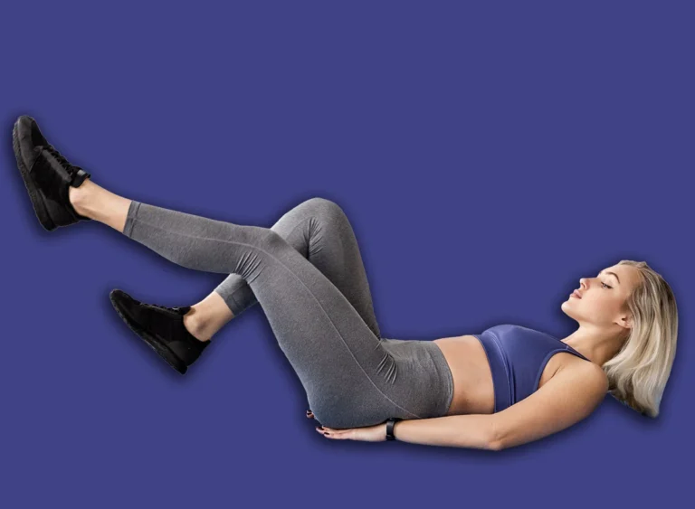
II. Revitalizing the Adductor Complex for Medial Stability
Another frequently assessed and critical component of hip health is the adductor complex, a group of muscles located on the inner thigh. The adductors are indispensable because they form a robust medial stabilizing chain that integrates directly with the pubic bone, the pelvic floor musculature, and the deeper layers of the abdominal system. Furthermore, many adductor muscles extend their attachments below the knee, rendering them key contributors to the stability of the entire lower limb.
When these adductor muscles lose their segmental functional capacity, the femoral head often struggles to maintain its optimal centration within the acetabular socket. This loss of precise centering can lead to either increased compressive loading or excessive laxity within the hip capsule, both of which are detrimental to joint health. Consequently, the manner in which forces are distributed through the knee and the sacroiliac (SI) region becomes significantly altered, often manifesting as pain or instability in these areas. Therefore, the strategic strengthening of the adductors transcends the simple action of squeezing the legs together; it is fundamentally about restoring balanced medial hip tension and re-establishing the harmonious fascial relationship between the pelvis and the trunk. This balanced tension is vital for proper joint mechanics and effective load transfer throughout the lower kinetic chain.
III. Enhancing Gluteus Maximus Activation and Myofascial Integrity
The gluteus maximus stands as one of the human body’s most substantial and powerful muscles, yet its multifaceted role is often underestimated. This muscle possesses diverse fiber orientations that connect directly to the sacrum, the expansive thoracolumbar fascia, the femur, and the iliotibial band (IT band). From a relational anatomical perspective, the gluteus maximus acts as a primary conduit for transmitting forces between the lower body and the spinal column, playing a pivotal role in movements like walking, running, and lifting.
When there is a decline in the segmental activation of the gluteus maximus, individuals commonly exhibit compensatory patterns where other muscles, such as the hamstrings or lumbar extensors, attempt to assume its responsibilities. This substitution directly shifts excessive mechanical load onto the lower back, often leading to altered pelvic rotation dynamics during daily activities. Restoring the precise segmental recruitment of the gluteus maximus is therefore essential for re-establishing appropriate posterior fascial tension and significantly enhancing the efficiency of load transfer through the pelvis into the spine.
Beyond mere strength, the myofascial quality of the gluteus maximus is equally critical. This muscle serves as a substantial fascial hub, interconnecting the sacrum, pelvis, and thoracolumbar fascia, and even exerting indirect influence on the mechanics of the contralateral shoulder via intricate fascial sling systems. Should this tissue become densified—meaning it loses its natural pliability and becomes stiff or restricted—it can severely impede proper pelvic rotation and diminish hip extension capabilities. This restriction, in turn, often translates into heightened tension throughout the lumbar spine. Implementing targeted myofascial stretching techniques is crucial for restoring the vital hydration and elasticity within these fascial layers, enabling the gluteus maximus to perform optimally as both a powerful stabilizer and an efficient force transmitter, thereby alleviating undue stress on the spine.
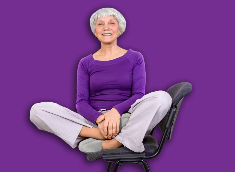
IV. Decompressing the Anterior Hip Capsule
A frequently overlooked yet critically important component in the hip-spine relationship is the anterior hip capsule. Over extended periods, particularly in the context of modern sedentary lifestyles involving prolonged sitting, the structures comprising the anterior coxofemoral joint can become chronically compressed, leading to a significant reduction in their inherent elasticity.
When the anterior capsule loses its physiological mobility and flexibility, it can physically alter the precise axis of rotation of the femoral head within the acetabulum. This seemingly minor alteration has profound implications that extend beyond mere hip mobility. It disrupts the proprioceptive input—the body’s sense of its position and movement—from the joint, which subsequently disorganizes how the surrounding stabilizing musculature functions and coordinates. In this context, specialized techniques like ELDOA (Global Osteoarticular Decoaptation Stretching) become immensely valuable. These methods are specifically designed to gently decompress the joint, meticulously restore the intricate balance of capsular tension, and profoundly enhance neurological awareness around the coxofemoral joint. By addressing this often-neglected area, the entire hip mechanism can function more harmoniously, contributing to overall stability and reducing referred pain.
In conclusion, the strategies outlined above represent an integrated approach to optimizing hip function, moving beyond isolated muscle strengthening to embrace the complex interplay of the entire joint system. By focusing on restoring precise hip centration, rebalancing the critical fascial load transfer pathways, and refining the neuromuscular coordination around the coxofemoral joint, the objective is to create a more resilient and efficient foundation for movement. When this intricate hip-spine relationship is systematically improved, the knees and lumbar spine almost invariably experience a significant reduction in chronic stress and discomfort. This holistic perspective offers a potent pathway for individuals to not only alleviate existing pain but also to proactively foster enduring spinal and lower extremity health, enabling a more active and fulfilling life in their maturing years.
About the Author


