
A groundbreaking advancement in medical imaging, developed by a collaborative team from the California Institute of Technology (Caltech) and the University of Southern California (USC), promises to revolutionize diagnostic capabilities by generating high-resolution, three-dimensional color visualizations of internal human tissues and their intricate vascular networks. This novel technique seamlessly integrates the visualization of anatomical structures with the real-time assessment of blood flow, offering unprecedented insight into physiological processes. Initial applications have already demonstrated its potential across various anatomical regions, with researchers highlighting its transformative impact on areas such as breast cancer detection, the nuanced tracking of nerve damage associated with diabetes, and the in-depth exploration of brain function.
The scientific community has detailed the intricacies of this innovative approach in a recent publication within the esteemed journal Nature Biomedical Engineering, signaling a significant step forward in the field of medical diagnostics.
Traditional medical imaging modalities, while indispensable, often present inherent limitations that this new technique aims to surmount. Conventional ultrasound, for instance, is lauded for its speed, affordability, and widespread availability, making it a staple in clinical settings. However, its primary strength lies in generating two-dimensional representations of tissue morphology, offering a comparatively restricted field of view and limited detail regarding subsurface structures. In parallel, photoacoustic imaging, a technique that leverages the interaction of laser light with biological tissues, provides a distinct form of information. This method involves directing laser pulses into the body and subsequently detecting the generated ultrasonic waves, which are emitted by molecules that absorb this light energy. This absorption process allows for the visualization of blood vessels in what can be described as optical color, and enables the observation of blood flow dynamics within arteries and veins. Nevertheless, photoacoustic imaging, on its own, struggles to capture the fine details of tissue architecture with the same clarity as other methods.
Other widely adopted imaging technologies, such as computed tomography (CT) and magnetic resonance imaging (MRI), each come with their own set of compromises. CT scans, while providing detailed cross-sectional images, involve the use of ionizing radiation, which necessitates careful consideration regarding patient exposure. MRI, on the other hand, offers exceptional soft tissue contrast and can visualize anatomical structures without radiation, but it often requires the administration of contrast agents, can be time-consuming, and is significantly more expensive to operate and utilize for frequent examinations. These trade-offs highlight a persistent need for imaging solutions that can offer a more comprehensive suite of information without introducing undue risks or escalating costs.
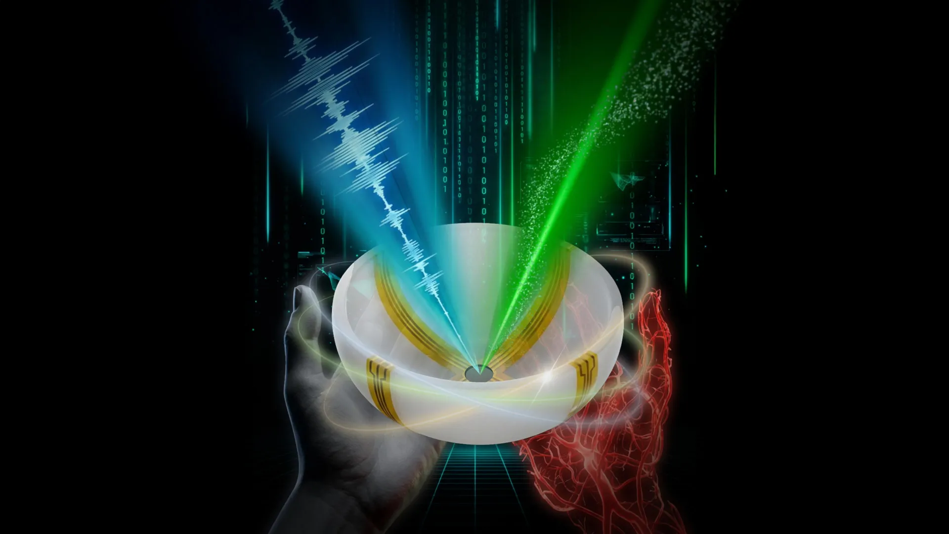
The research team’s innovative solution, christened RUS-PAT (Rotational Ultrasound Tomography combined with Photoacoustic Tomography), represents a sophisticated fusion of these complementary imaging principles. The foundational concept of photoacoustic tomography itself boasts a rich history, having been pioneered over two decades ago by Professor Lihong Wang, a distinguished figure in medical engineering and electrical engineering at Caltech. In its original form, PAT operates by employing short laser pulses that illuminate biological tissues. Molecules within these tissues that absorb the light energy undergo rapid thermal expansion, generating acoustic waves. These acoustically emitted signals are then detected and processed to reconstruct detailed images, effectively mapping out the distribution of light-absorbing chromophores, such as hemoglobin in blood.
Professor Wang articulated the primary objective of this new project: to synergistically combine the inherent strengths of both ultrasound and photoacoustic imaging modalities. He emphasized that this was not a simple additive process, but rather a complex endeavor requiring the development of an optimized integration strategy. The challenge lay in devising a system that could leverage the structural imaging capabilities of ultrasound with the functional and molecular information provided by photoacoustic techniques in a cohesive and efficient manner.
Addressing the complexities and costs associated with traditional imaging systems, particularly ultrasound, the researchers sought a more streamlined and practical design. Conventional ultrasound apparatuses typically employ an array of numerous individual transducers to both emit and receive sound waves. Integrating such a multi-transducer setup directly with the requirements of photoacoustic imaging presented significant engineering hurdles, rendering it overly complicated and prohibitively expensive for widespread clinical adoption. Photoacoustic imaging, in contrast, primarily relies on ultrasound detectors for signal acquisition. This disparity in hardware requirements sparked a novel conceptual leap for Professor Wang. He pondered the possibility of adapting the photoacoustic principle by reversing the excitation mechanism. Instead of relying on laser light to generate ultrasound waves, he envisioned utilizing ultrasound itself to mimic the excitation process, thereby creating a system that could capture signals from both modalities using a shared detection apparatus.
The breakthrough came with the realization that a single, broad-area ultrasound transducer could be employed to transmit sound waves that propagate throughout the tissue. Crucially, the same detector array could then be utilized to capture the acoustic signals generated by both the direct ultrasound transmission and the photoacoustic effect. This elegant solution bypassed the need for separate, complex transducer arrays for each modality.
The finalized system incorporates a relatively small number of arc-shaped ultrasound detectors. These detectors are strategically arranged and designed to rotate around a central point. This rotational configuration effectively simulates a full hemispheric detection coverage, providing comprehensive data acquisition while maintaining a significantly simpler and more cost-effective hardware architecture compared to previous integrated approaches. This design simplification is a critical factor in paving the way for broader accessibility and implementation in clinical settings.
The demonstrated potential for human application of this innovative imaging technique has been met with considerable enthusiasm from the medical and scientific communities. Dr. Charles Y. Liu, a co-author of the study and a distinguished researcher and clinician at USC, underscored the significance of this development. He noted that the novel amalgamation of acoustic and photoacoustic methodologies effectively addresses many of the persistent limitations encountered with existing, widely utilized medical imaging tools in current clinical practice. Furthermore, he emphasized the critical validation of the technique’s feasibility for human use, evidenced by its successful application in multiple anatomical contexts during the study. Dr. Liu, who holds professorial appointments at the Keck School of Medicine of USC and directs USC’s Neurorestoration Center, also serves as the chair of neurosurgery at the Rancho Los Amigos National Rehabilitation Center, bringing a wealth of clinical perspective to the research.
The inherent versatility of the RUS-PAT method, particularly its ability to function wherever light can penetrate, suggests a broad spectrum of potential clinical applications. In the realm of breast cancer diagnostics, for example, this technology could empower clinicians to not only precisely pinpoint the location of a tumor but also to glean crucial information regarding its underlying biological activity and metabolic status. For individuals suffering from diabetic neuropathy, a condition characterized by nerve damage often accompanied by impaired blood flow, the technique offers the unprecedented capability to simultaneously assess both the structural integrity of the nerves and the oxygen supply to these tissues within a single, non-invasive scan. Professor Wang further elaborated on its promising role in neuroscience research, where it could enable scientists to conduct simultaneous investigations of brain anatomy and dynamic blood flow patterns, thereby deepening our understanding of neurological processes and diseases.
Key performance metrics of the RUS-PAT system, including its speed and imaging depth, have also been subject to rigorous evaluation. At its current stage of development, the system is capable of imaging tissue up to approximately 4 centimeters in depth. The researchers are also exploring avenues to extend its reach into deeper anatomical regions. The delivery of light for photoacoustic excitation can be achieved through the use of endoscopic tools, which hold the potential to access areas of the body not readily accessible by external illumination. A significant advantage of this new modality is its remarkable speed; each RUS-PAT scan can be completed in under one minute, a considerable improvement over many existing time-consuming imaging procedures.
The physical configuration of the current experimental setup involves positioning ultrasound transducers and a laser source beneath a specialized scanning bed, designed to accommodate patients comfortably during the imaging process. The system has undergone successful preliminary testing with human volunteers and patients, and is currently progressing through the initial phases of its transition toward broader clinical implementation. This progression signifies a crucial step in translating cutting-edge research into tangible benefits for patient care.
The comprehensive scientific report detailing this research is titled "Rotational ultrasound and photoacoustic tomography of the human body." The study’s co-lead authors include Yang Zhang and Shuai Na, who conducted their foundational work as postdoctoral researchers at Caltech and are now affiliated with Tsinghua University and Peking University, respectively. Dr. Jonathan J. Russin, also a co-lead author, contributes his expertise from his affiliations with the Keck School of Medicine of USC and the Rancho Los Amigos National Rehabilitation Center in Downey, California.
A cadre of additional researchers from Caltech played pivotal roles in the development of this technology. These contributors include Karteekeya Sastry, Li Lin (who completed her PhD in 2020 and is now at Zhejiang University in Hangzhou, China), Junfu Zheng, Yilin Luo, Xin Tong (who earned her Master of Science in 2021), Yujin An, Peng Hu (who completed his PhD in 2023), and former research scientist Konstantin Maslov. Dr. Tze-Woei Tan from the Keck School of Medicine of USC is also acknowledged as a co-author, further underscoring the inter-institutional collaboration. The foundational research and development of this transformative imaging modality were made possible through generous funding from the National Institutes of Health, a testament to the critical support provided by government agencies for pioneering scientific endeavors.
About the Author


