

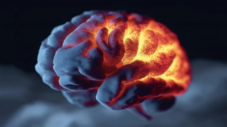
Alzheimer’s disease casts a long, devastating shadow over millions globally, progressively stripping individuals of their cognitive faculties, most notably their capacity for memory. This neurodegenerative condition is characterized by an insidious decay of brain cells and the intricate neural connections that underpin thought, emotion, and recollection. While the catastrophic end-stage effects are well-documented, the precise molecular mechanisms initiating this cellular destruction have remained a complex and contentious frontier in neuroscience. For decades, a dominant theory has centered on the accumulation of amyloid-beta, a protein fragment that forms plaques in the brain, widely believed to be a primary driver of neuronal damage. However, the multifaceted nature of Alzheimer’s has also implicated a host of other biological factors, including the entanglement of tau proteins, dysfunction of cellular waste-disposal systems known as lysosomes, chronic neuroinflammation, and the aberrant activity of immune cells like microglia.
A groundbreaking investigation, recently published in the esteemed Proceedings of the National Academy of Sciences, offers a significant step toward unifying some of these disparate theories. Researchers have unveiled compelling new evidence suggesting that two of the most prominent hypotheses regarding Alzheimer’s progression—the amyloid-beta cascade and inflammatory responses—may converge upon a singular molecular pathway. This critical convergence point involves a specific receptor that appears to instruct neurons to dismantle their synapses, the vital communication junctions between brain cells. This discovery not only provides a potential missing link between previously distinct lines of inquiry but also challenges long-held assumptions about how memory loss unfolds in the disease.
The pivotal research was spearheaded by Dr. Carla Shatz, a distinguished affiliate of the Wu Tsai Neurosciences Institute and the Sapp Family Provostial Professor, alongside Dr. Barbara Brott, a research scientist within Dr. Shatz’s laboratory. Their collaborative efforts received crucial financial backing, including a Catalyst Award from the Knight Initiative for Brain Resilience, an ambitious program dedicated to re-evaluating the fundamental biological underpinnings of neurodegenerative disorders like Alzheimer’s. This support underscores the growing recognition of the need for innovative approaches to understanding and combating these complex conditions.
Central to the study’s findings is a protein receptor identified as LilrB2. Dr. Shatz’s extensive work on this molecule spans many years, establishing a profound understanding of its physiological roles. As early as 2006, her team made a seminal discovery, revealing that the mouse homolog of LilrB2 plays an indispensable role in synaptic pruning—a naturally occurring process essential for refining neural circuits during brain development and for facilitating learning and memory formation throughout adulthood. Synaptic pruning, in its healthy manifestation, is akin to a sculptor refining their work, removing excess or inefficient connections to strengthen crucial pathways.
The significance of LilrB2 to Alzheimer’s pathology began to crystallize with subsequent research. In 2013, Dr. Shatz’s group demonstrated that amyloid-beta, the notorious protein fragment associated with Alzheimer’s plaques, could directly bind to LilrB2. This binding event was shown to act as a trigger, initiating a cascade within neurons that led to the removal of synapses. Crucially, further experimental manipulations revealed that genetically removing this receptor in mouse models of Alzheimer’s disease provided significant protection against the characteristic memory deficits, strongly implicating LilrB2 as a key mediator of pathological synapse loss. These earlier findings laid the groundwork for the more recent, broader insights into the receptor’s role.
Parallel to the amyloid-beta research, another major focus of the study explored the intricate workings of the immune system, specifically a process known as the complement cascade. This sophisticated system is a vital component of the body’s innate immunity, responsible for releasing molecules that aid in the identification and elimination of pathogens such as viruses and bacteria, as well as damaged cells. Under normal conditions, the complement cascade is a guardian of cellular health. However, chronic inflammation has long been recognized as a significant risk factor for Alzheimer’s disease, creating a fertile ground for pathological processes.
Mounting evidence from recent studies has increasingly linked dysregulation of the complement cascade to excessive, aberrant synaptic pruning and to the progression of various neurological disorders. These observations prompted Dr. Shatz and her team to hypothesize whether molecules involved in inflammatory responses might interact with the LilrB2 receptor in a manner analogous to amyloid-beta, thereby contributing to the pathological loss of synapses. This line of reasoning suggested a potential convergence point where immune system dysregulation could directly impact neural connectivity.
To rigorously test this novel hypothesis, the research team embarked on a systematic screening of various molecules within the complement cascade, meticulously searching for any that could bind to the LilrB2 receptor. Their exhaustive investigation yielded a singular, compelling candidate: the protein fragment C4d. This particular molecule demonstrated a sufficiently strong attachment to LilrB2 to suggest its direct involvement in mediating synapse loss. The identification of C4d as a binding partner for LilrB2 represented a critical breakthrough, bridging the inflammatory response with a known synaptic pruning mechanism.
To validate this in vitro finding, the researchers moved to in vivo experiments, testing their hypothesis in living organisms. They carefully injected C4d into the brains of healthy mice and observed the resultant effects. The outcome was striking and, as Dr. Shatz noted, quite surprising, especially given that C4d had previously been thought to be a functionally inert molecule. "Lo and behold," she recounted, "it stripped synapses off neurons." This direct demonstration of C4d’s capacity to induce synapse loss in a living brain provided powerful confirmation of its active role and solidified the connection between inflammation, the complement cascade, and synaptic destruction.
When these comprehensive findings are considered in their totality, they strongly indicate that both amyloid-beta accumulation and inflammatory processes may drive the pathological loss of synapses through the very same biological mechanism, specifically via the LilrB2 receptor. This profound insight necessitates a fundamental re-evaluation of current models explaining how Alzheimer’s disease progressively erases memories. The traditional view of separate, parallel pathways contributing to neurodegeneration may need to be updated to incorporate this shared, downstream effector.
"There’s an entire set of molecules and pathways that lead from inflammation to synapse loss that may not have received the attention they deserve," remarked Dr. Shatz, who also holds professorships in biology within the School of Humanities and Sciences and in neurobiology within the School of Medicine. Her statement underscores the potential for an entirely new avenue of research and therapeutic intervention, moving beyond the well-trodden paths of amyloid and tau.
Furthermore, these results directly challenge a long-standing assumption prevalent within Alzheimer’s research concerning the primary agents of synaptic removal. For many years, it was widely believed that glial cells—the brain’s immune and support cells, such as microglia and astrocytes—were predominantly responsible for the pathological elimination of synapses during disease progression. However, the current study suggests a more direct and active role for neurons themselves in this destructive process. "Neurons aren’t innocent bystanders," Dr. Shatz asserted. "They are active participants." This paradigm shift implies that the very cells responsible for transmitting information are also, under pathological conditions, actively involved in dismantling their own communication networks, adding a crucial layer of complexity to the disease’s pathogenesis.
This newfound understanding carries significant implications for the future development of Alzheimer’s therapies. Current FDA-approved treatments for the disease primarily focus on reducing amyloid plaques in the brain. While these drugs have offered some benefits, they have often been accompanied by limited efficacy and significant side effects. Dr. Shatz pointed out the inherent challenges: "Busting up amyloid plaques hasn’t worked that well, and there are a lot of side effects," including concerning issues such as headaches and brain bleeding. She further emphasized that "even if they worked well, you’re only going to solve part of the problem." This perspective highlights the need for strategies that address multiple facets of the disease, moving beyond a singular focus on amyloid clearance.
The research suggests that a more efficacious therapeutic strategy might involve directly targeting receptors like LilrB2, which directly orchestrate synapse removal. By developing interventions that protect these crucial synaptic connections, Dr. Shatz posits, it may be possible to safeguard memory itself from the ravages of Alzheimer’s. Such an approach would represent a shift from merely attempting to clear pathological protein aggregates to actively preserving the functional integrity of neural circuits—a potentially more direct route to preserving cognitive function.
The extensive study involved a collaborative team of researchers, including Barbara Brott, Aram Raissi, Monique Mendes, Caroline Baccus, Jolie Huang, and Carla Shatz from Stanford University’s Department of Biology, Stanford Medicine’s Department of Neurobiology, and Bio-X; Kristina Micheva from Stanford’s Department of Molecular and Cellular Physiology; and Jost Vielmetter from the California Institute of Technology. This interdisciplinary effort underscores the complex nature of modern scientific discovery. Generous funding support was provided by multiple esteemed organizations, including the National Institutes of Health, the Sapp Family Foundation, the Champalimaud Foundation, the Harold and Leila Y. Mathers Charitable Foundation, the Ruth K. Broad Biomedical Research Foundation, and the Phil and Penny Knight Initiative for Brain Resilience at the Wu Tsai Neuroscience Institute at Stanford University. The critical provision of human Alzheimer’s disease tissue samples by the Neurodegenerative Disease Brain Bank at the University of California, San Francisco, further supported by NIH funding and other consortia, was instrumental in allowing the researchers to validate their findings in human-relevant contexts, underscoring the collaborative spirit essential for advancing our understanding of this challenging disease.
About the Author


