Unveiling the Role of Lipid-Engorged Immune Cells in Aggravating Multiple Sclerosis Pathology
A groundbreaking investigation spearheaded by Daan van der Vliet and his collaborators at the Netherlands Institute for Neuroscience, in conjunction with Leiden University and Utrecht University, has illuminated a crucial biological mechanism that may elucidate the heightened severity of multiple sclerosis (MS) observed in certain individuals. By meticulously examining neural tissue harvested from patients exhibiting rapid MS progression, the research team identified a significant prevalence of atypical immune cells characterized by an abundance of intracellular fat droplets. These findings hold considerable promise for the development of novel therapeutic interventions and the identification of future diagnostic indicators capable of forecasting disease trajectory.
Multiple sclerosis is fundamentally a neurodegenerative condition characterized by the progressive deterioration of myelin, the lipid-rich sheath that insulates nerve fibers within the central nervous system, encompassing the brain and spinal cord. This insulation is indispensable for the efficient transmission of neural signals, and its breakdown invariably leads to a spectrum of neurological deficits, ranging from motor impairments like difficulty with ambulation to visual disturbances. The inherent heterogeneity of MS progression presents a persistent challenge for the medical community, with some patients experiencing a protracted and relatively mild clinical course spanning many years, while others face a rapid onset of severe disability, including paralysis, even at a young age. Deciphering the underlying factors that contribute to this disparity in outcomes has been a paramount objective for scientific inquiry into MS.
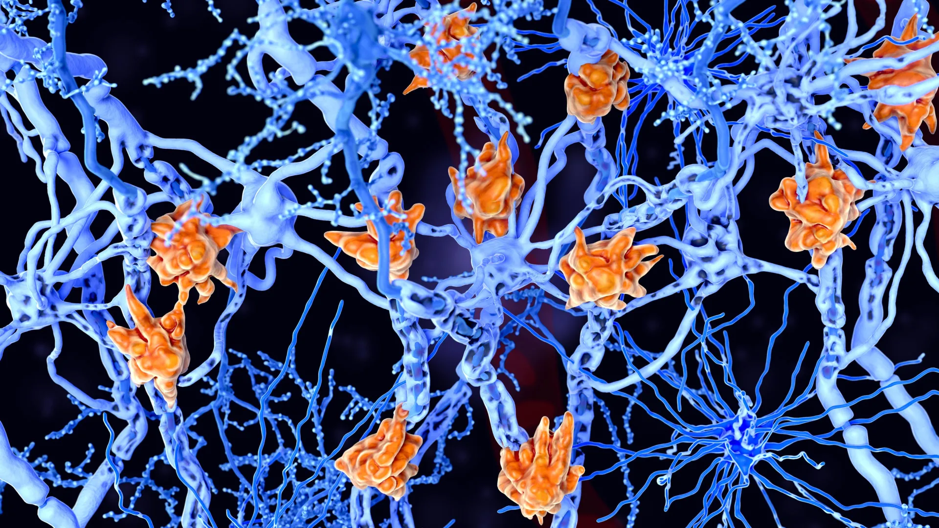
The research endeavors were specifically directed towards understanding the behavior of microglia, the resident immune cells of the central nervous system, which normally perform vital functions such as clearing cellular debris and facilitating tissue repair. However, in the context of MS, these cells undergo a profound transformation, becoming engorged with lipid particles, thereby acquiring a distinctive "foamy" appearance. Scientists have termed these altered cells "foamy microglia."
"We observed a consistent correlation between a higher density of these foamy microglia and a more aggressive disease progression in patients," stated lead researcher Daan van der Vliet, underscoring the significance of this cellular phenotype.
Normally, microglia act as vigilant guardians of brain health, diligently removing damaged cellular components and waste products. In the pathological landscape of MS, it is hypothesized that these microglia inadvertently become overwhelmed by the sheer volume of myelin debris they attempt to clear. This excessive accumulation of lipids exceeds their metabolic processing capacity, leading to their characteristic foamy transformation.
"The prevailing theory is that these cells are initially attempting a beneficial function: the removal of pathological material," Van der Vliet elaborated. "However, they become critically overloaded, compromising their ability to effectively contribute to the intricate process of neural repair."
Further granular analysis of the MS lesions revealed distinct molecular signatures differentiating areas characterized by the presence of foamy microglia from those that were devoid of them. Specifically, regions populated by foamy microglia exhibited an elevated concentration of certain lipid species that have been historically associated with sustained inflammatory activity.
The long-standing understanding of MS pathogenesis has predominantly implicated inflammation as the primary driver of disease advancement. Nevertheless, the current research findings introduce a more nuanced perspective, suggesting that the disease trajectory may involve a more intricate cascade of events than previously appreciated.
"The pathology appears to extend beyond a simple overreliance on inflammatory responses alone," Van der Vliet commented. "It seems these cells are engaged in a failing attempt to clear damage and promote healing, which paradoxically exacerbates inflammation and hinders recovery."
This perspective highlights a critical aspect of the disease mechanism: a biological process initially designed for protective and reparative functions can, under pathological conditions, become a contributing factor to ongoing neural damage when its normal operational capacity is compromised.
The scientific foundation for these revelations was built upon the meticulous examination of post-mortem human brain tissue obtained from 28 individuals diagnosed with MS, who had generously bequeathed their brains for scientific research through the Netherlands Brain Bank.
Employing a sophisticated array of cutting-edge analytical techniques in parallel, the researchers were able to simultaneously assess gene expression patterns, protein profiles, and lipid compositions within individual MS lesions. This integrated approach facilitated the construction of a highly detailed and comprehensive molecular portrait of the biological processes unfolding in the affected brain regions.
Van der Vliet emphasized the synergistic interplay between advanced technological capabilities and a deep-seated understanding of neuropathology as being indispensable for the successful execution of this research.
"Contemporary scientific tools offer unprecedented precision in mapping the intricate architecture of the brain," Van der Vliet noted. "However, the interpretative power of these technologies is significantly amplified when they can be directly correlated with observable pathological changes in neural tissue. The enduring legacy of meticulous study and classification of brain tissue by the Netherlands Brain Bank provided the essential framework for recognizing these aberrant cellular patterns."
The implications of this discovery extend significantly towards the potential for developing more precise prognostic tools and personalized treatment strategies for MS patients. The research identified preliminary evidence suggesting that specific lipids associated with foamy microglia might also be detectable in cerebrospinal fluid. If corroborated by subsequent investigations, these molecular markers could serve as valuable biomarkers, enabling the identification of individuals at a higher risk of experiencing rapid disease deterioration.
"This opens up exciting avenues for the future development of biomarkers that could assist clinicians in earlier identification of patients predisposed to rapid decline, thereby guiding them towards the most appropriate therapeutic interventions," Van der Vliet explained.
Furthermore, these findings align seamlessly with ongoing efforts to develop novel therapeutic agents that specifically target lipid metabolism pathways and the expansion of chronic MS lesions. Several experimental treatment modalities are presently undergoing evaluation in clinical trials, often conducted in collaboration with pharmaceutical partners such as Roche, further underscoring the translational potential of this research.
The investigation was generously supported by two prestigious Gravitation programs: the Institute for Chemical Immunology (ICI) and the Institute for Chemical NeuroScience (iCNS), underscoring the collaborative and well-funded nature of this significant scientific endeavor.
Related posts:

Related News