
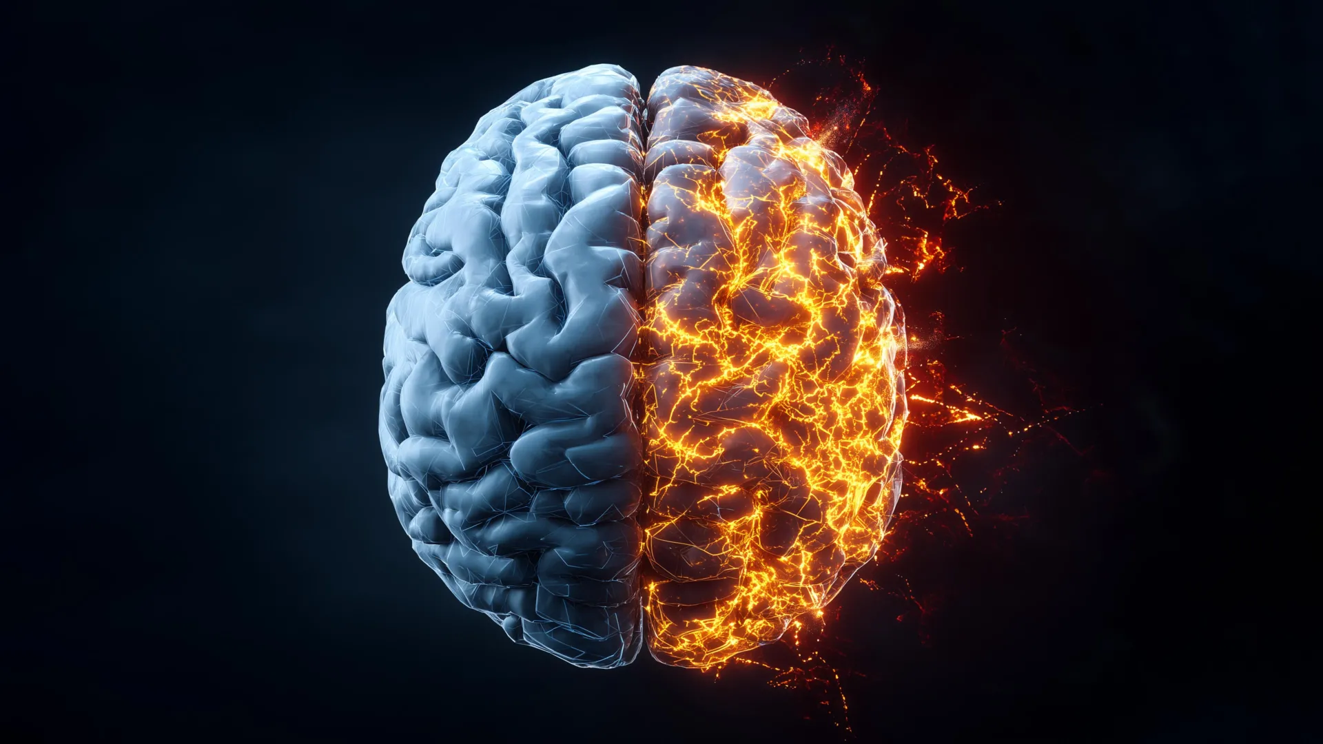
Researchers at Rice University have pioneered the development of the inaugural comprehensive, label-free molecular atlas of the Alzheimer’s-affected brain within an animal model, offering an unprecedentedly granular perspective on the disease’s genesis and propagation. The profound impact of Alzheimer’s, which tragically claims more lives annually than breast and prostate cancers combined, underscores the critical imperative for a deeper comprehension of its underlying drivers. This groundbreaking study, detailed in the latest issue of ACS Applied Materials and Interfaces, leverages a sophisticated fusion of advanced light-based imaging techniques and artificial intelligence to reveal that the chemical alterations characteristic of Alzheimer’s disease extend far beyond the well-documented amyloid plaques. Instead, these molecular disturbances manifest throughout the brain’s intricate architecture in complex, heterogeneous patterns, challenging existing paradigms of disease understanding.
The scientific team employed a highly refined form of Raman spectroscopy, known as hyperspectral Raman imaging, to meticulously detect these subtle yet significant chemical shifts within brain tissue. This advanced methodology utilizes a precisely controlled laser to elicit and capture the unique molecular signatures, or "fingerprints," inherent to the various chemical compounds present in biological samples. Ziyang Wang, a doctoral candidate in electrical and computer engineering at Rice and a lead author on the research, explained the system’s efficacy: "Traditional Raman spectroscopy provides a single data point regarding chemical information at a specific molecular location. In contrast, hyperspectral Raman imaging performs this measurement repeatedly, thousands of times across an entire tissue section, thereby constructing a comprehensive molecular map. The resultant output is an exquisitely detailed visualization illustrating the spatial variations in chemical composition across diverse regions of the brain."
The researchers systematically scanned entire brain sections, meticulously accumulating a vast repository of overlapping measurements to construct high-resolution molecular atlases for both healthy and Alzheimer’s-diseased tissues. A key feature of this innovative approach is its "label-free" nature, meaning the biological samples were examined in their native state, without the introduction of exogenous dyes, fluorescent proteins, or molecular tags that could potentially alter or obscure the natural chemical landscape. "This enabled us to observe the brain’s inherent chemical makeup, capturing an unadulterated and complete portrait of its molecular composition," Wang remarked. "We believe this unbiased methodology is more adept at uncovering novel disease-related changes that might otherwise remain undetected."
The sheer volume of data generated by this high-throughput imaging process necessitated the application of advanced machine learning (ML) algorithms for comprehensive analysis. Initially, the researchers employed unsupervised ML techniques, allowing sophisticated algorithms to identify inherent patterns within the spectral data without any preconceived notions or prior assumptions about disease markers. These models were thus able to categorize and cluster different tissue regions based purely on their unique molecular characteristics. Subsequently, the team transitioned to supervised ML, where algorithms were trained on datasets comprising both Alzheimer’s-affected and healthy brain samples. This training phase enabled the models to accurately distinguish between the two conditions and quantify the degree to which specific brain regions exhibited Alzheimer’s-related chemical signatures.
The findings from this analytical phase revealed a striking heterogeneity in the disease’s impact: "We observed that the chemical alterations induced by Alzheimer’s disease are not uniformly distributed throughout the brain," stated Wang. "Certain areas display pronounced chemical modifications, while others remain comparatively less affected. This uneven distribution pattern offers a potential explanation for the gradual onset of symptoms and may shed light on why therapeutic strategies targeting a single pathological mechanism have historically yielded limited success."
Beyond the accumulation of aberrant proteins, the study unearthed broader metabolic discrepancies between healthy and Alzheimer’s-burdened brains. Specifically, regional variations in cholesterol and glycogen levels were identified, with the most significant disparities occurring in brain regions critically involved in memory formation and retention, namely the hippocampus and the cerebral cortex. Shengxi Huang, an associate professor of electrical and computer engineering and materials science and nanoengineering, and the corresponding author of the study, elaborated on the significance of these metabolic findings. "Cholesterol plays a vital role in maintaining the structural integrity of brain cells, and glycogen serves as a readily accessible local energy reserve," Professor Huang explained. "Collectively, these observations lend robust support to the growing understanding that Alzheimer’s pathology encompasses more pervasive disruptions in brain structure and energy homeostasis, extending beyond mere protein aggregation and misfolding." Professor Huang’s extensive affiliations include the Ken Kennedy Institute, the Rice Advanced Materials Institute, and the Smalley-Curl Institute, highlighting the interdisciplinary nature of this research.
The genesis of this ambitious project stemmed from ongoing dialogues among researchers seeking innovative methodologies for studying the complex biological processes occurring within the Alzheimer’s-affected brain. Initially, the researchers were focused on analyzing only localized regions of brain tissue. However, Wang conceived of a more expansive approach: "I began to consider the potential of mapping the entire brain to gain a significantly broader perspective," he recalled. "Achieving this required numerous iterations of testing and refinement through trial and error before the imaging measurements and subsequent data analysis converged into a cohesive and functional workflow."
Upon the successful compilation of the complete molecular map, the impact of the findings was immediate and profound. "Patterns emerged that had been entirely invisible with conventional imaging techniques," Wang reported, expressing his deep satisfaction. "Witnessing those results was incredibly gratifying. It felt akin to uncovering a hidden stratum of information that had been present all along, awaiting the appropriate analytical tools to be revealed." By providing the first detailed, dye-free chemical maps of the Alzheimer’s brain, this pioneering research offers a more holistic and nuanced understanding of the disease’s progression. The research team anticipates that these insights will ultimately pave the way for earlier and more accurate diagnostic capabilities, as well as the development of more effective therapeutic interventions aimed at slowing the relentless advancement of Alzheimer’s disease. The foundational work for this research was generously supported by grants from the National Science Foundation (awards 2246564 and 1934977), the National Institutes of Health (award 1R01AG077016), and the Welch Foundation (award C2144).
About the Author


