A groundbreaking investigation spearheaded by researchers at Stanford Medicine is fundamentally reshaping our comprehension of brain development, challenging the long-held scientific consensus that the human brain originates from a singular developmental lineage. This pioneering work suggests that what we perceive as a unified cerebral organ may, in fact, be a sophisticated amalgamation of two distinct neural systems, each tracing its evolutionary roots back hundreds of millions of years. This revelation promises to unlock new avenues for understanding complex neurological disorders and refine our approaches to regenerative medicine.
For an extended period, the prevailing wisdom in neuroscience has posited the brain as a singular, cohesive organ, tracing its entire development back to a uniform cellular precursor. Under this widely accepted doctrine, the various specialized regions—the forebrain, responsible for higher cognitive functions; the midbrain, involved in motor control and sensory information; and the hindbrain, which governs essential life-sustaining processes—were all thought to derive from the same initial pool of progenitor cells during embryonic growth. This monolithic view underpinned much of the research and theoretical frameworks concerning brain formation and function.
However, the recently published findings, detailed in the prestigious journal Nature Neuroscience, paint a dramatically different picture. The Stanford team’s meticulous research indicates that the human brain integrates two ancient and separately evolving nervous systems. One of these primordial systems gives rise to the structures indispensable for fundamental physiological operations, such as the rhythmic regulation of breathing and the consistent pumping of the heart. The other distinct system produces the neural circuitry associated with sophisticated human capabilities, including the intricate processes of language acquisition, advanced mathematical reasoning, abstract thought, and the unique capacity for self-reflection and introspection. This bifurcation at the earliest stages of development represents a significant departure from previous models.
This novel insight carries profound implications, potentially resolving a long-standing conundrum within the neuroscientific community: the persistent difficulty in successfully cultivating certain types of brain cells, particularly those from the hindbrain region, within laboratory settings. The previous struggles, the research suggests, stemmed from an incorrect understanding of the cells’ developmental origins. Furthermore, this discovery opens an entirely new frontier for investigating debilitating diseases that specifically affect the brain stem, such as spinal muscular atrophy (SMA), a devastating genetic condition primarily affecting infants, and amyotrophic lateral sclerosis (ALS), also known as Lou Gehrig’s disease, a progressive neurodegenerative ailment impacting motor neurons.
Dr. Kyle Loh, an associate professor of developmental biology and the senior author of the study, emphasized the significance of their findings: "We have definitively demonstrated for the first time that the anterior regions of the brain emerge from a progenitor cell population entirely distinct from that which forms the posterior brain structures." He added, "Our breakthrough fundamentally alters our ability to generate and study hindbrain neurons in a controlled environment, such as a petri dish, allowing us to scrutinize their functions with unprecedented precision." This newfound capability promises to accelerate research into diseases where these specific neurons are compromised.
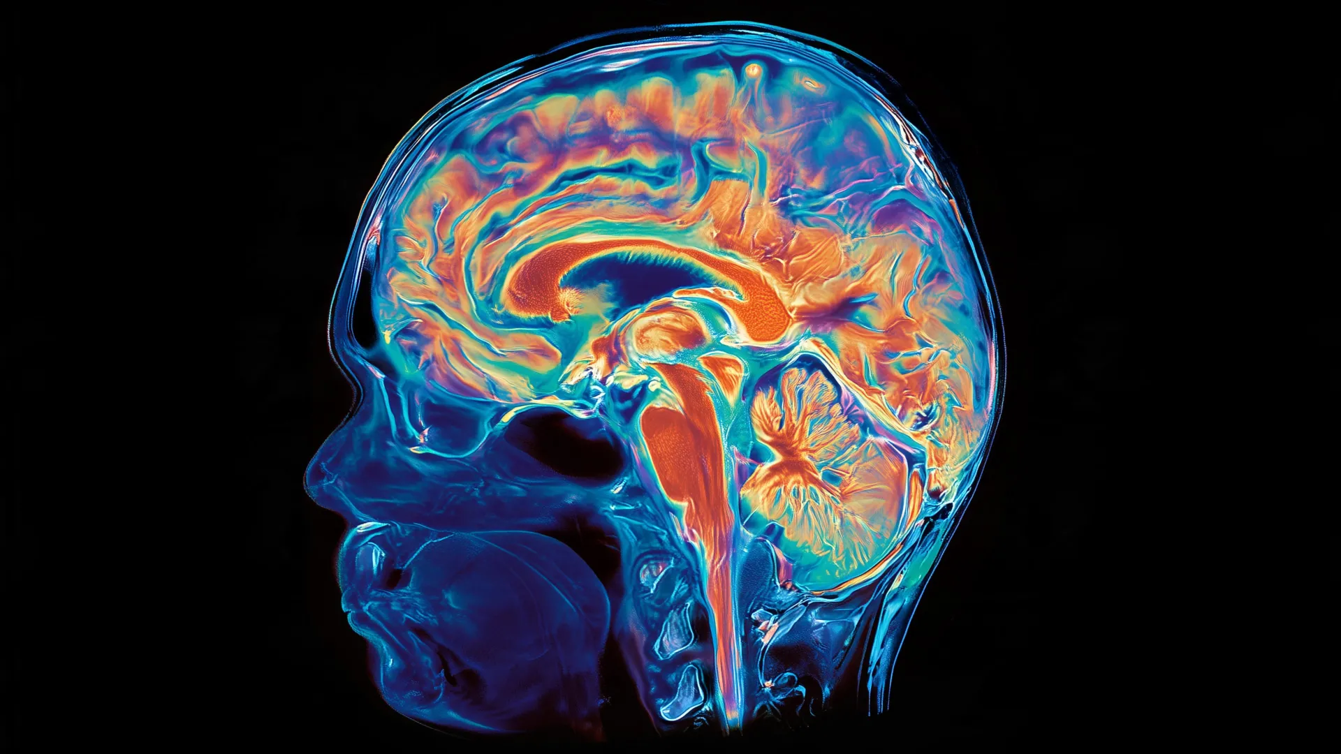
The adult human brain is conventionally segmented into three principal divisions. The forebrain, situated at the front, underpins many of the faculties most closely associated with advanced cognition, encompassing linguistic processing, conscious awareness, and abstract analytical reasoning. Conversely, the hindbrain, often referred to as the brain stem, is located at the posterior base of the skull. It is the command center for automatic, life-sustaining functions, including the regulation of respiration, sleep cycles, cardiac rhythm, and the sensation of hunger. Beyond these autonomic controls, hindbrain neurons also orchestrate the complex movements of facial muscles, the tongue, and the throat, which are crucial for articulation and deglutition (swallowing).
Despite the hindbrain’s indispensable role in survival and essential functions, neuroscientists have faced decades of frustration in their attempts to engineer human hindbrain neurons in laboratory cultures. This significant technical limitation has historically impeded comprehensive research into severe neurological conditions that specifically target the brain stem. Spinal muscular atrophy, for instance, remains a leading genetic contributor to mortality in children under the age of one. Amyotrophic lateral sclerosis, typically diagnosed between the ages of 40 and 70, impacts both forebrain and hindbrain neurons. In both SMA and ALS, a progressive deterioration of particular hindbrain neurons occurs, leading to a gradual loss of vital functions. Patients may eventually lose the ability to swallow, significantly increasing the risk of aspiration pneumonia if food or liquids inadvertently enter the lungs. In advanced stages, the ability to breathe independently can also be compromised.
The pivotal insight that illuminated this dual developmental pathway emerged from the researchers’ meticulous examination of the earliest phases of embryonic development, specifically during a critical process known as gastrulation. Gastrulation is a fundamental stage where the single-layered blastula reorganizes into a multi-layered structure, establishing the basic body plan and laying down the groundwork for all future tissues and organs. It was during this nascent period that graduate students Carolyn Dundes and Rayyan Jokhai, co-first authors of the research, made their crucial observation.
They discovered that the hindbrain does not arise as a subsequent branch or a downstream differentiation of the developmental trajectory that produces the forebrain and midbrain. Instead, the hindbrain embarks upon its own independent developmental pathway right from the outset, progressing in parallel with the formation of the other major brain regions. To meticulously trace this intricate process, the research team employed developing mouse embryos, a well-established model for human development. Through careful observation, they successfully identified two distinct populations of brain progenitor cells.
One population of these progenitor cells was characterized by the expression of a specific gene, Otx2, and was definitively destined to form the forebrain and midbrain. A second, entirely separate population of progenitor cells expressed a different gene, Gbx2, and was committed to developing into the hindbrain. Crucially, the researchers observed that these two distinct cell populations remained spatially separate and did not intermingle or overlap, even at the very earliest stages of embryonic development they were able to examine.
This fundamental distinction became even more pronounced when the team delved into the organization of chromatin, the complex material comprising DNA and proteins that condenses to form chromosomes within the nucleus of eukaryotic cells. Chromatin plays a critical role in regulating gene expression, dictating which genes are accessible for transcription and which remain silenced. The anterior neural ectoderm, which forms the future forebrain and midbrain, exhibited fundamentally different chromatin configurations compared to the posterior neural ectoderm, which gives rise to the future hindbrain. These inherent differences in chromatin structure effectively "locked in" the cells to their separate developmental fates from the very beginning.
Rayyan Jokhai elaborated on the implications of this finding for past research efforts: "Previous attempts to generate hindbrain neurons in the lab likely tried to coerce forebrain and midbrain progenitors into adopting a hindbrain identity, which our study unequivocally demonstrates is biologically impossible." This insight offers a compelling explanation for the persistent challenges encountered by earlier researchers. They may have been attempting to force a cell type into a developmental pathway that it was never programmed to undertake. Jokhai further emphasized the importance of their approach, stating, "In the field of stem cell biology, there’s often a singular focus on achieving the final, differentiated cell type, such as a mature neuron. However, it is paramount to meticulously investigate the earliest stages of embryonic development. Our careful attention to this nascent time point allowed us to uncover this fundamental divergence in brain formation."
Armed with this unprecedented understanding of the hindbrain’s independent developmental trajectory, the research team was able to overcome a significant technical hurdle in laboratory science. For the first time, they successfully guided human pluripotent stem cells—remarkable cells capable of differentiating into virtually any cell type in the human body—to mature into functional hindbrain motor neurons. These laboratory-grown cells exhibited authentic characteristics of genuine hindbrain neurons, generating electrical signals known as action potentials and producing specific proteins associated with regions of the hindbrain that control facial and swallowing muscles. This breakthrough promises an inexhaustible supply of relevant cells for future research.
The researchers then expanded their investigation to explore the evolutionary depth of this developmental split. Spanning over 550 million years of evolutionary history, the team unearthed compelling evidence of the same two-origin arrangement in a diverse array of species, including chickens, zebrafish, and even acorn worms. Acorn worms are small, marine invertebrates that dwell on the seafloor and represent a very distant common ancestor shared with humans, making their developmental similarities particularly striking. The team also observed that jellyfish, whose evolutionary lineage diverged from that of humans approximately 600 to 700 million years ago, possess two distinct nervous systems positioned at opposite ends of their bodies.
Collectively, these findings strongly suggest the profound possibility that the modern vertebrate brain did not evolve as a single, unitary structure, but rather arose when evolutionary pressures led to the spatial convergence and integration of two pre-existing, ancient neural systems. Dr. Loh mused on this evolutionary ingenuity: "Our research posits that evolution effectively brought two existing neural systems into close physical proximity, merging them spatially. While having the brain as a singular, contiguous organ might seem more efficient, we appear to rely on this ancient, primordial mechanism to construct the brain as two separate, yet interconnected, components." Jokhai echoed this sentiment, expressing his astonishment: "I was genuinely surprised by our findings because the very word ‘brain’ implies a unified, contiguous organ with a singular origin. Yet, even half a billion years ago, these separate neural systems existed, and now they operate almost as one, which is truly remarkable."
This groundbreaking work holds immense potential for scientists dedicated to studying and ultimately combating SMA, ALS, and other devastating disorders that compromise the brain stem. A major impediment in this field has been the limited access to viable human tissue. Obtaining brain stem samples from living patients for routine research is generally not feasible, making direct examination of affected neurons exceptionally challenging. The newfound ability to cultivate functional hindbrain neurons in a laboratory dish dramatically alters this landscape, empowering scientists to investigate the underlying pathological mechanisms of these diseases with unprecedented precision and detail.
Beyond neurodegenerative diseases, the hindbrain also plays a crucial role in another significant area of medical research: weight management. It harbors intricate neural circuits that regulate hunger and satiety, systems that are directly influenced by contemporary weight-loss medications such as semaglutide. This broader involvement underscores the extensive medical relevance of the hindbrain and the potential for this research to impact diverse fields.
Looking ahead, the research team is now focused on further elucidating the developmental origins of the spinal cord and meticulously determining how SMA and ALS specifically disrupt the delicate functions of hindbrain neurons. "We now possess a robust model to gain a deeper understanding of these devastating diseases," Jokhai concluded, "and crucially, to work towards developing effective regenerative therapies for them. This truly marks a very exciting new frontier in brain research." The collaboration involved researchers from the California Institute of Technology and the University of California, San Francisco, with funding from numerous prestigious institutions, including the National Institutes of Health, the National Science Foundation, and several private foundations.
About the Author