
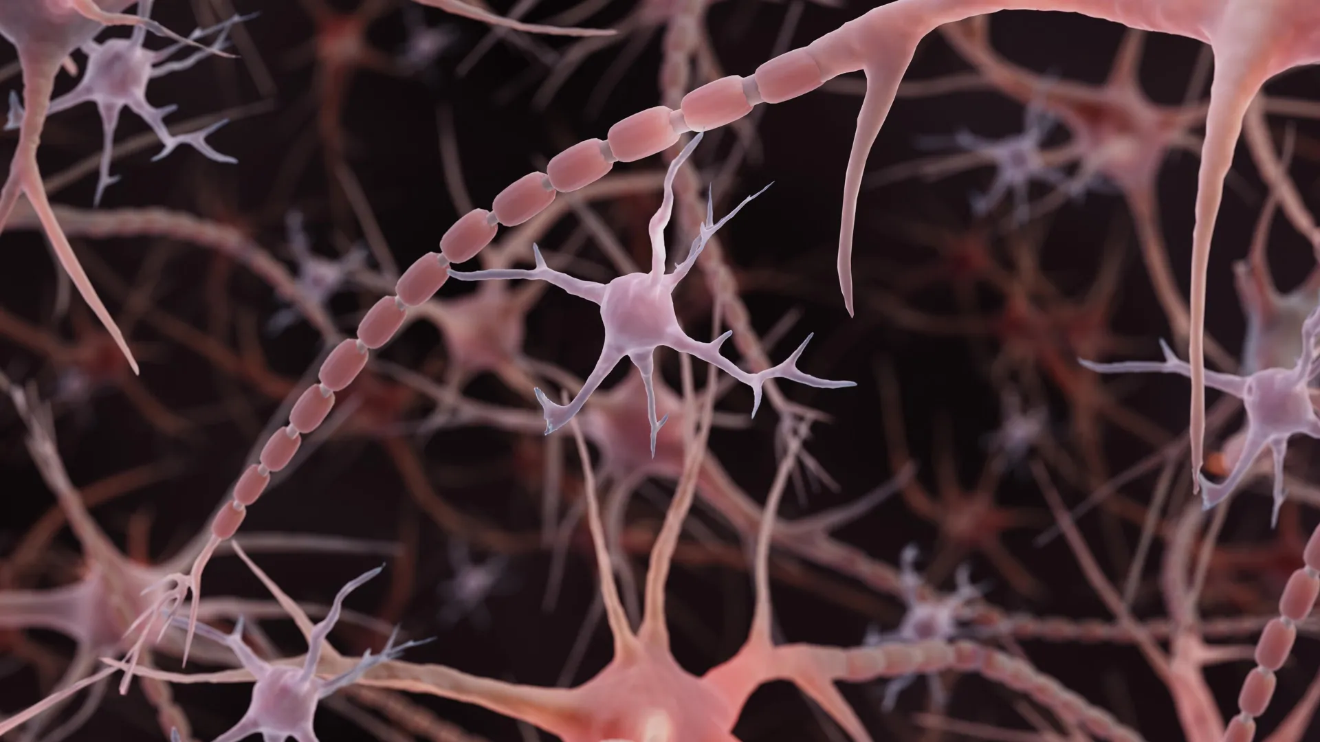
For over a century, the prevailing depiction of nerve cell extensions, known as axons, in educational materials has presented them as smooth, unvarying conduits responsible for transmitting electrical signals throughout the brain. However, groundbreaking research is compelling a significant revision of this fundamental neuroanatomical understanding, revealing that these critical communication lines may possess a far more intricate and dynamic structure than previously assumed. Instead of the familiar, uniform tubular form, scientists have discovered that many axons exhibit a distinct "beaded" or "pearled" appearance, characterized by repeating, nanoscale swellings along their length. This discovery, first meticulously detailed in a study published in Nature Neuroscience in late 2024 by researchers at Johns Hopkins Medicine, suggests that the physical morphology of axons plays a more influential role in regulating neural communication than has been appreciated.
The conventional textbook illustration of an axon typically portrays a slender tube, occasionally punctuated by larger bulges known as synaptic varicosities. These varicosities are well-understood to be crucial sites for the storage and release of neurotransmitters, the chemical messengers that facilitate signal transmission between neurons. While the presence of prominent swellings along axons has been observed in contexts of cellular distress, such as during neuronal death or in neurodegenerative conditions like Parkinson’s disease, where such beading is often associated with damage to the cell membrane and its internal structural scaffolding, the recent findings highlight a different phenomenon. The Johns Hopkins team identified much smaller, regularly spaced swellings that appear to be a feature of healthy, functioning axons. They have termed these structures "non-synaptic varicosities," distinguishing them from the known communication-related bulges and suggesting that this patterned morphology might represent an inherent aspect of normal axonal architecture.
The genesis of this line of inquiry can be traced to earlier observations of similar bead-like formations along axons in simpler organisms, specifically worms, by Shigeki Watanabe, Ph.D., an associate professor of cell biology and neuroscience at the Johns Hopkins University School of Medicine. These initial observations, coupled with discussions with Swiss scientist Graham Knott, Ph.D., spurred a deeper investigation into the underlying causes of this peculiar axonal structure. One hypothesis considered the role of the axon’s internal cytoskeleton, a network of protein filaments that provides structural support and helps maintain the cell’s shape. Previous research had identified repeating structural elements within axons, leading Watanabe and Knott to explore whether disrupting this internal framework might cause the beaded appearance to disappear. However, experiments conducted by Jacqueline Griswold, the study’s lead author and a graduate student at Johns Hopkins, demonstrated that the pearling persisted even when the internal skeletal components were manipulated, redirecting the focus toward the physical properties of the axon’s outer membrane.
This shift in perspective led to a collaborative effort involving theoretical biophysicist Padmini Rangamani, Ph.D., a professor at the University of California San Diego School of Medicine, to explore how the surrounding cell membrane might actively shape the axon’s morphology. To meticulously examine these microscopic structures, which are approximately 100 times narrower than a human hair, the researchers employed a cutting-edge imaging technique: high-pressure freezing electron microscopy. This method is crucial because traditional electron microscopy often involves chemical fixation and dehydration, processes that can inadvertently alter the very structures they aim to observe. By contrast, high-pressure freezing rapidly solidifies the cellular sample, preserving its delicate architecture in a state much closer to its natural condition, akin to preserving a grape’s form rather than reducing it to a raisin through dehydration.
The study meticulously analyzed axons from various sources, including mouse neurons cultured in the laboratory, neurons from adult mice, and those derived from mouse embryos. Importantly, the researchers focused on nonmyelinated axons, which lack the insulating myelin sheath that typically encases longer axons in the central nervous system. Across tens of thousands of detailed images, the researchers consistently observed the characteristic pearled or beaded structure. Furthermore, the presence of these swellings was confirmed using high-resolution imaging of living neurons, providing compelling evidence that the observed morphology was not an artifact of the sample preparation process itself. Watanabe remarked that these findings fundamentally challenge a century of established understanding regarding axon structure.
To elucidate the mechanisms responsible for this recurring beaded pattern, the research team developed mathematical models that simulated the physical behavior of the axon’s surrounding membrane. These relatively simple mechanical models proved adept at reproducing key features of the observed swellings, suggesting that physical forces acting on the membrane could be sufficient to dictate axonal shape, without necessarily relying on a rigid internal structural blueprint. Experimental validation further supported this interpretation. For instance, altering the osmotic environment by increasing the sugar concentration in the surrounding solution led to a reduction in the size of the swollen regions. In parallel, the mathematical models indicated that increased membrane tension also correlated with smaller swellings.
The role of cholesterol within the cell membrane was also investigated as a means to modulate its mechanical properties. By reducing cholesterol levels, the membrane became less rigid and more fluid. This alteration in membrane fluidity, observed in both the computational models and the actual mouse neurons, directly impacted the beaded structure and, notably, resulted in a deceleration of electrical signal propagation along the axon. Watanabe explained that while wider axonal spaces might intuitively suggest unimpeded ion flow, the interplay between the size of the swellings and the narrower connecting segments plays a critical role in determining the overall efficiency of signal transmission. Intriguingly, the study revealed that larger swellings did not always equate to faster signals; the precise dimensions of both the bulges and the constrictions between them were significant factors.
Beyond static structural observations, the researchers also investigated whether the axons’ physical form could dynamically respond to neural activity. Following periods of high-frequency electrical stimulation, the pearled regions exhibited notable changes, increasing in length by an average of 8% and widening by 17%. This structural alteration persisted for at least 30 minutes post-stimulation. Crucially, the electrical signals transmitted along these stimulated axons slowed down, an effect that lasted for at least an hour. When cholesterol was depleted prior to stimulation, some structural modifications still occurred, but the swellings remained smaller than those in untreated cells, and the significant signal slowing was not observed, further underscoring the interplay between membrane composition, structure, and electrical signaling. These findings strongly suggest that axons are not merely passive, fixed conduits but rather possess a remarkable capacity to physically adapt in response to neural activity, thereby fine-tuning the efficiency of information transfer.
The implications of these findings were soon extended to human biology when a subsequent study, involving co-authorship by Watanabe, examined axonal morphology in human brain tissue. This related research, published in Neuron in late 2025, investigated cortical tissue obtained from individuals undergoing epilepsy surgery. By combining electrical stimulation with rapid freezing techniques, similar to those used in the initial mouse studies, the researchers captured subtle membrane changes in both mouse and human brain slices. Alongside the identification of pearled axons in human tissue, the study provided evidence of ultrafast endocytosis, a rapid membrane retrieval process occurring at nerve endings after the release of chemical signals. While these observations broaden the scope of the findings to include human physiology, they do not definitively establish that all axons share this beaded structure or that the effects on signaling are uniform across the entire human brain. The human study primarily focused on membrane recycling dynamics at synapses.
The fundamental question of what controls axonal shape and its relationship to function has garnered significant attention and support. Watanabe and Rangamani were awarded a National Institute of Mental Health (NIMH) Multiple Principal Investigator grant, highlighted in 2024, to further explore this area. This project aims to develop sophisticated computational models predicting how a neuron’s intrinsic physical properties and incoming neural signals influence its axonal morphology, with subsequent experimental validation in mouse tissue and cell cultures. Concurrently, other research groups are developing novel methodologies to probe the intricate dimensions of neural wiring. For instance, a study published in PLOS Biology in July 2026 utilized automated imaging of living zebrafish to screen a large library of compounds for their effects on axonal diameter. This research identified several compounds that influence specific chemical signaling pathways and can alter axon width, providing an additional valuable tool for understanding the regulatory mechanisms governing axon size, even if not directly replicating the pearling phenomenon.
For the Johns Hopkins team and the broader neuroscience community, a critical ongoing question is how the newly characterized structure of healthy, pearled axons relates to the more pronounced, damaging beading observed in cases of neurological disease. Differentiating between these two forms of axonal morphology could prove instrumental in distinguishing between normal, adaptive changes in neural wiring and the pathological breakdown of neuronal function, potentially paving the way for more targeted diagnostic and therapeutic strategies.
About the Author












