Pioneering Research Unravels Primate Visual Acuity Genesis in Utero
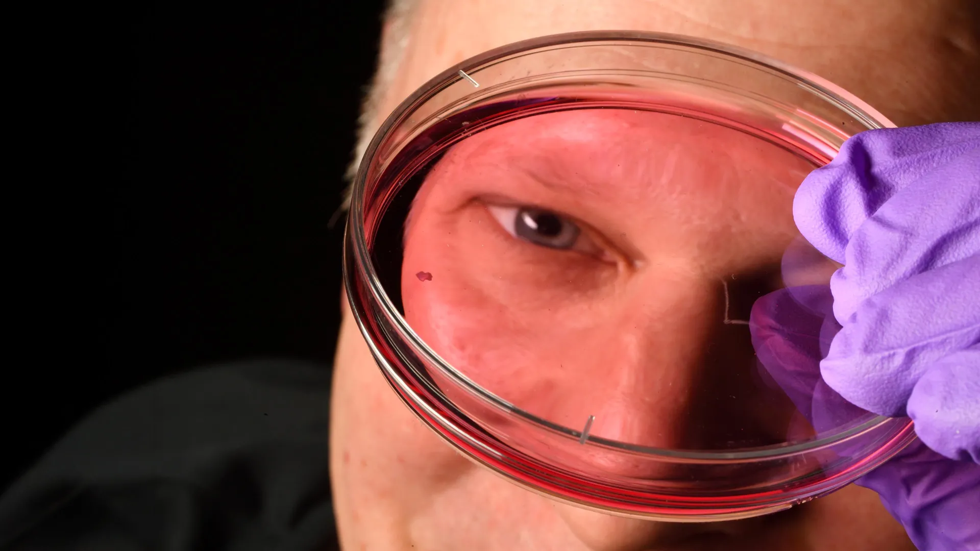
A groundbreaking revelation from Johns Hopkins University researchers has illuminated the intricate developmental pathway by which humans acquire their exceptional central visual acuity prior to birth, pinpointing a precisely orchestrated interplay between a vitamin A metabolite and thyroid hormones within the retinal tissue. This significant scientific advancement fundamentally challenges a long-held understanding of how crucial light-sensitive cells, known as photoreceptors, are established and holds considerable promise for the development of novel therapeutic strategies targeting a spectrum of debilitating vision impairments, including age-related macular degeneration, glaucoma, and other conditions that progressively degrade sight. The findings, derived from meticulous studies utilizing laboratory-cultivated retinal tissues, have been formally documented and disseminated in the prestigious journal Proceedings of the National Academy of Sciences.
At the heart of this transformative research lies the creation and prolonged observation of retinal organoids, miniature three-dimensional structures meticulously grown from embryonic stem cells, designed to faithfully replicate key aspects of the human retina’s architecture and function. These sophisticated bioengineered tissues, maintained and studied over several months, provided an unprecedented window into the complex cellular choreography that culminates in the formation of the foveola. This specialized, minute depression at the very center of the retina is the linchpin of our sharpest, most detailed vision, enabling us to discern fine textures, read small print, and recognize faces with remarkable clarity.
The investigation specifically zeroed in on the development of cone photoreceptors, the specialized cells responsible for our ability to perceive color and see with clarity in well-lit conditions. These cells, destined to differentiate into distinct types sensitive to different wavelengths of light – namely blue, green, and red – are paramount to the richness of our visual experience. While the foveola constitutes a minuscule fraction of the overall retinal surface area, it is disproportionately responsible for an estimated half of all visual information processed by the human brain. Intriguingly, the foveola exhibits a unique composition compared to the peripheral retina; it is predominantly populated by red and green cones, with blue cones conspicuously absent in the mature state.
For decades, the precise mechanisms governing this specialized arrangement of cone photoreceptors within the foveola has remained an enigmatic puzzle for vision scientists. A significant hurdle in unraveling this mystery has been the inherent biological differences between human vision development and that observed in commonly used laboratory model organisms, such as mice and fish, which do not possess the same intricate photoreceptor distribution. This species-specific divergence has limited the applicability of traditional animal models to fully elucidate the human visual system’s formation.
The newly published research proposes a paradigm shift in our understanding, suggesting that the distinctive cone cell pattern within the foveola emerges through a meticulously synchronized cascade of developmental events occurring early in fetal development. Specifically, between the 10th and 12th weeks of gestation, an initial population of blue cones transiently appears within the developing foveal region. However, by approximately the 14th week, a remarkable transformation takes place: these nascent blue cones undergo a phenotypic conversion, evolving into the red and green cones that characterize the mature foveola.
This profound cellular metamorphosis, the study reveals, is orchestrated by a dual-action molecular mechanism. Initially, a molecule derived from vitamin A, known as retinoic acid, plays a critical role in regulating the developmental trajectory. The researchers observed that the localized breakdown or modulation of retinoic acid signaling appears to curtail the generation of new blue cones. Following this initial regulatory step, thyroid hormones then assume a pivotal role, actively driving the existing population of blue cones to reprogram their cellular identity and differentiate into red and green cones.
Dr. Robert J. Johnston Jr., an associate professor of biology at Johns Hopkins University and the lead investigator of this pivotal study, elaborated on the significance of these findings. He emphasized that this discovery represents a critical advance toward comprehensively understanding the intricate biological processes underpinning the central retina, a region of paramount importance to vision and the primary site of degeneration in conditions like macular degeneration. "By gaining a deeper insight into this specific retinal area and developing organoids that accurately mimic its sophisticated functionality, our ultimate aspiration is to pave the way for the laboratory generation and subsequent transplantation of these tissues, thereby restoring lost vision," Dr. Johnston stated.
The research team’s findings directly challenge a prevailing scientific hypothesis that has guided vision research for approximately thirty years. The long-standing model posited that the few blue cones initially present in the foveal region were somehow actively displaced or migrated outwards, making way for the red and green cones. In stark contrast, the new evidence strongly supports an alternative model: that these cells do not emigrate but rather undergo an intrinsic transformation, changing their functional type from blue to red or green cones. This cellular plasticity, the researchers highlight, is a truly surprising revelation that reframes our understanding of developmental biology within the eye.
The implications of this research extend far beyond fundamental biological inquiry, offering a beacon of hope for the future of vision restoration therapies. The research team is actively engaged in refining their retinal organoid technology, aiming to create models that more closely recapitulate the complexity and functionality of the native human retina. The development of these enhanced models could significantly accelerate the production of healthier photoreceptor cells, which are essential for future cell replacement therapies. Such therapies hold immense potential for treating devastating diseases like macular degeneration, for which effective cures currently remain elusive.
This cutting-edge work is a testament to the power of advanced bioengineering and molecular biology in tackling complex biological questions. The scientific community anticipates that further exploration of these developmental pathways, coupled with advancements in regenerative medicine, could unlock novel therapeutic avenues for millions worldwide affected by vision loss. The long-term vision is to engineer "made-to-order" populations of photoreceptor cells that can be safely and effectively integrated into the damaged eye, potentially re-establishing functional vision and dramatically improving the quality of life for patients. While the journey toward clinical application is acknowledged to be extensive, requiring rigorous optimization and extensive safety studies, this research marks a significant and promising stride in that direction.

Related News