
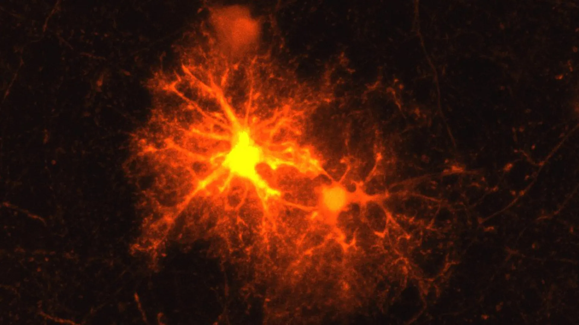
When the intricate architecture of the brain sustains damage, a remarkable cellular mobilization occurs, a phenomenon consistently observed in laboratory investigations involving rodent models. Dr. Jan Deussing, a seasoned neurobiologist and leader of a research group, has repeatedly witnessed the emergence of a specific cohort of cells congregating around injured neural tissue, their activity intensifying in response to the trauma. Despite numerous observations of this localized cellular response, the precise identity of these crucial players remained an enigma, presenting an ideal challenge for an aspiring researcher.
This compelling mystery became the central focus of Clemens Ries’s master’s thesis, undertaken during an internship at the Max Planck Institute of Psychiatry as he neared completion of his biology degree. Employing a meticulously designed mouse model, Ries embarked on a systematic process of elimination, testing a comprehensive array of cellular markers to pinpoint the elusive cell type. His exhaustive investigation yielded a singular, definitive result: the marker for oligodendrocyte progenitor cells (OPCs) uniquely illuminated the activated cells in the vicinity of neural injury.
OPCs represent a vital class of precursor cells within the central nervous system, possessing the remarkable capacity to differentiate into oligodendrocytes. These mature cells are the architects of the myelin sheath, a fatty insulating layer that encases the axons of nerve cells. Axons, the slender extensions of neurons, serve as the communication conduits, enabling electrochemical signals to traverse the vast neural network. Myelin’s function is analogous to the protective insulation surrounding an electrical wire, facilitating efficient signal transmission and providing essential metabolic support to the axons. This insulating layer is thus fundamental to the optimal functioning of the brain.
Disruptions to the myelin sheath can precipitate a cascade of severe neurological consequences. In the context of autoimmune disorders such as multiple sclerosis (MS), the body’s own immune system mistakenly attacks and degrades this protective coating. Physical trauma to the brain can also inflict damage upon myelin, and in instances of significant injury, the resulting degradation can even lead to the demise of entire neurons. Consequently, the restoration of myelin around compromised axons is a critical component of the brain’s inherent capacity for repair following injury.
Ries’s initial exploration of these newly identified cells for his master’s project quickly blossomed into a passion, compelling him to continue his investigation into a doctoral thesis. His subsequent research unearthed a previously unrecognized aspect of the brain’s response to injury: the dramatic proliferation of OPCs at the periphery of damaged brain tissue. Crucially, a significant proportion of these OPCs then embark on a developmental trajectory, maturing into fully functional oligodendrocytes capable of synthesizing new myelin.
However, the groundbreaking discoveries of Ries and Deussing extended beyond the mere identification and quantification of these repair cells. They uncovered a surprising biochemical signaling event: a substantial subset of OPCs, approximately one-third, located near the injured site, were observed to produce and release corticotropin-releasing hormone (CRH). CRH is a neuropeptide widely recognized for its pivotal role in mediating the body’s physiological response to stress. This finding marked a significant departure from established knowledge, as researchers had not previously understood that OPCs possessed the capability to synthesize neuropeptides like CRH. The implications of these findings, now formally documented in the prestigious journal Cell Reports, are profound.
The initiation of the CRH response following an injury is remarkably swift, with detectable production occurring within mere hours of the insult. This surge in CRH signaling, however, proves to be transient, subsiding after approximately three days. This brief but potent burst strongly suggests that CRH plays a crucial, time-sensitive role during the earliest phases of the brain’s healing process.
Central to this intricate regulatory mechanism appears to be one of the two known receptors for CRH, specifically CRH receptor 1 (CRHR1). This receptor is expressed on a distinct population of OPCs, enabling them to detect and respond to the CRH released by their activated counterparts. Experimental investigations revealed a compelling correlation: when CRHR1 is absent, OPCs exhibit a more rapid rate of proliferation in the aftermath of an injury. Paradoxically, this accelerated initial expansion does not translate into more effective myelin repair. Instead, the ultimate outcome is a reduced number of mature oligodendrocytes, compromising the integrity of the repaired myelin sheath.
These observations collectively indicate that CRH acts as a critical regulator, dictating the precise timing of OPC maturation. This temporal control appears to be paramount for ensuring the generation of a sufficient population of mature oligodendrocytes, a prerequisite for the effective restoration of damaged myelin.
The significance of OPCs extends beyond their role in post-injury repair; they are also integral to the process of myelination during normal brain development. This developmental myelination, a crucial period for establishing neural circuitry, commences in utero and continues well into young adulthood. Given that CRHR1 is present on OPCs even in the absence of injury, Ries and Deussing hypothesized that this receptor might also influence myelination during the brain’s natural developmental trajectory. Collaborating with other research teams, they employed a battery of experimental methods to investigate myelin formation in additional mouse models.
Their research demonstrated that mice genetically engineered to lack CRHR1 exhibited an increased proliferation of OPCs during the initial stages of development. These developmental alterations were not transient; they persisted and exerted lasting effects on the structural organization of the brain. In adult brains of these mice, researchers observed significant changes in myelination patterns, characterized by the formation of thicker myelin sheaths, particularly around thinner axons. These findings strongly suggest that CRHR1 on OPCs plays a dual role, not only in facilitating myelin repair after injury but also in orchestrating the fundamental processes of myelin development from its inception.
The question of CRH’s origin during normal brain development presents a distinct inquiry compared to its post-injury surge. While injured OPCs themselves act as a source of CRH, the source of this crucial hormone during developmental myelination remains to be definitively elucidated. The scientific team proposes a compelling hypothesis: that developing neurons may be the primary source of CRH during this critical period. Their model posits that as neurons mature, they release CRH, which in turn influences both the proliferation of OPCs and their subsequent differentiation into myelin-producing oligodendrocytes.
This proposed mechanism carries significant implications when considered in the broader context of neurological and psychiatric health. Neurons are already known to release CRH, particularly under conditions of stress. Furthermore, early-life stress is a well-established risk factor for the development of psychiatric disorders. The newly uncovered role of CRH in OPC regulation and myelin development raises the tantalizing possibility that the CRH system operating within these progenitor cells may have far-reaching consequences for mental well-being.
Dr. Deussing speculates that the CRH system within OPCs might play a more substantial role than previously understood in stress-associated psychiatric conditions, such as depression. Should future research endeavors corroborate and expand upon this potential link, a deeper comprehension of how CRH signaling influences OPC behavior, myelin formation, and overall brain development could pave the way for the development of entirely novel therapeutic strategies aimed at addressing a spectrum of neurological and psychiatric challenges.
About the Author












