
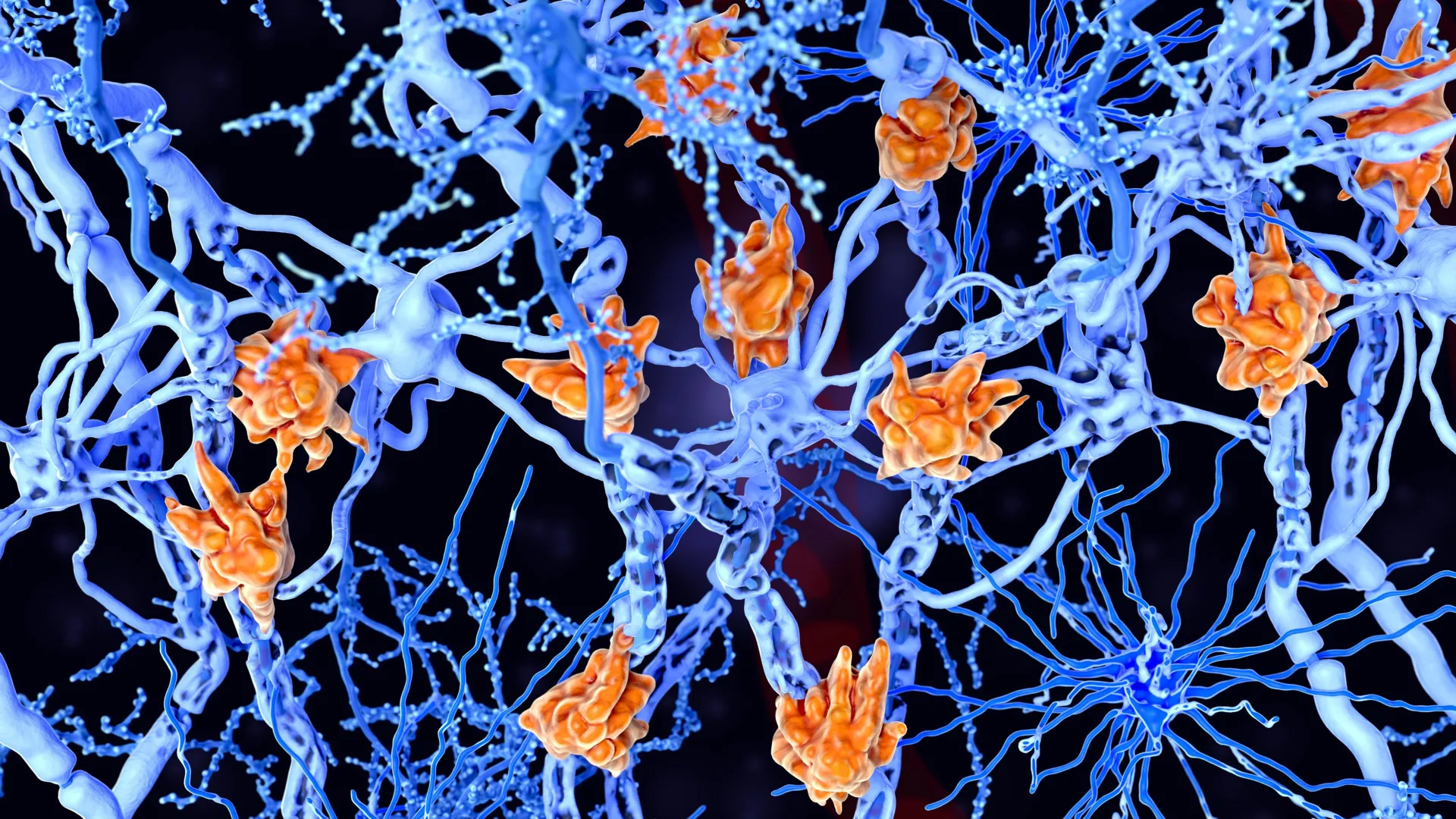
A groundbreaking study conducted by an international consortium of neuroscientists has unveiled a critical cellular mechanism that may elucidate why certain individuals experience a more aggressive trajectory with multiple sclerosis (MS). The research, spearheaded by Daan van der Vliet and involving collaborative efforts from the Netherlands Institute for Neuroscience, Leiden University, and Utrecht University, meticulously examined post-mortem brain tissue from patients diagnosed with rapidly advancing MS. Their findings revealed an unexpectedly high prevalence of specialized immune cells within the brain, identified as microglia, which were engorged with lipid droplets, giving them a characteristic "foamy" appearance. This discovery holds significant promise for the development of novel therapeutic interventions and the identification of predictive biomarkers to anticipate disease severity and rate of decline.
Multiple sclerosis is a chronic, autoimmune disease that profoundly impacts the central nervous system, specifically targeting myelin. Myelin, a fatty sheath, acts as an essential insulator for nerve fibers, facilitating rapid and efficient transmission of electrochemical signals. In MS, the immune system mistakenly attacks and degrades this vital myelin layer, a process known as demyelination. The ensuing disruption of nerve signal conduction leads to a diverse array of neurological deficits, often manifesting as motor impairments, sensory disturbances, visual problems, and cognitive challenges. The heterogeneity of MS progression, ranging from benign, slowly evolving forms to severe, debilitating conditions characterized by rapid disability and paralysis, has long been a central enigma in neurological research, prompting continuous investigation into the underlying biological drivers of this variability.
At the heart of this recent investigation are microglia, the resident immune cells of the brain. Their primary roles encompass surveillance of the neural environment, clearance of cellular debris, and the orchestration of tissue repair processes. However, under pathological conditions such as MS, microglia can undergo significant morphological and functional alterations. The researchers observed that in the context of advanced MS, these crucial cells accumulate substantial quantities of lipids, derived from damaged myelin, leading to their distended, foamy morphology. This transformation suggests a deviation from their intended supportive functions, potentially contributing to disease exacerbation.
"We observed a strong correlation between the presence of a significant number of these lipid-laden microglia and a more severe clinical presentation of the disease," stated lead researcher Daan van der Vliet, underscoring the pivotal nature of their observation. This finding directly links a specific cellular phenotype to disease severity, offering a tangible target for further inquiry.
The study posits that these foamy microglia represent an overwhelmed system attempting to cope with overwhelming cellular damage. Normally, microglia are adept at phagocytosing, or engulfing, cellular debris, including fragments of damaged myelin, to facilitate tissue cleanup and promote healing. In the relentless onslaught of MS, however, it is theorized that microglia may ingest myelin breakdown products at a rate that exceeds their metabolic capacity for processing and clearance. This overload results in the intracellular accumulation of lipids, transforming the microglia into their foamy state and, critically, impairing their ability to effectively contribute to restorative processes.
"The prevailing hypothesis is that these cells are engaged in an earnest effort to perform their essential duty of clearing pathological material," explained Van der Vliet. "However, they become functionally incapacitated due to this excessive lipid burden. Consequently, their capacity to support neural repair is compromised, and they may inadvertently exacerbate the ongoing pathology."
Further molecular analyses performed by the research team revealed significant disparities between MS lesions characterized by the presence of foamy microglia and those lacking them. Specifically, areas heavily populated by these foamy cells exhibited a marked enrichment of certain lipid species. These particular lipids are known to be associated with persistent inflammatory activity, suggesting a complex interplay where the cellular response to damage might, paradoxically, sustain and amplify inflammation.
Historically, inflammation has been considered a primary driver of MS pathogenesis. However, this new research introduces a more nuanced perspective, proposing a cascade of events where the initial attempt at cellular cleanup by microglia, when it fails, can fuel a cycle of chronic inflammation and hinder recovery. The findings suggest that a mechanism initially designed for neuroprotection can, under pathological duress, transform into a contributor to sustained neural damage. "It is becoming increasingly clear that the disease process is not solely attributable to the direct inflammatory response," Van der Vliet elaborated. "Instead, it appears to involve a complex feedback loop where the microglia’s failed attempt to manage myelin debris leads to a worsening of the inflammatory environment and a suppression of the body’s natural repair mechanisms."
The research team meticulously analyzed brain tissue samples from 28 individuals who had bequeathed their brains to the Netherlands Brain Bank for scientific study following their diagnosis of MS. This invaluable resource provided the foundation for their detailed investigation. Employing a suite of cutting-edge analytical techniques concurrently, the scientists were able to simultaneously assess gene expression patterns, protein profiles, and lipid composition within individual MS lesions. This multi-omic approach enabled the construction of an exceptionally detailed molecular and cellular map of the affected brain regions, offering unprecedented insight into the pathological processes at play.
The successful integration of advanced technological capabilities with a deep understanding of neuropathology was deemed critical to the study’s success. "Modern scientific instrumentation offers remarkable precision in mapping the intricate details of the brain," Van der Vliet remarked. "However, the true value of these technologies is unlocked when their findings can be directly correlated with the observable pathology in human brain tissue. The extensive, decades-long meticulous classification and study of brain tissue conducted by the Netherlands Brain Bank provided the essential context for recognizing and interpreting these aberrant cellular patterns."
The implications of this discovery extend significantly towards the future of personalized MS management. The identification of foamy microglia and their associated lipid signatures could pave the way for the development of novel diagnostic tools and therapeutic strategies. The researchers found preliminary evidence suggesting that certain fats linked to the foamy microglia phenotype might be detectable in the cerebrospinal fluid. If corroborated by further studies, these molecules could serve as invaluable biomarkers, enabling clinicians to more accurately identify patients at elevated risk of rapid disease progression at an earlier stage.
"This opens up exciting avenues for the development of predictive biomarkers that could assist clinicians in identifying individuals prone to rapid neurological decline, thereby facilitating the selection of the most appropriate and effective therapeutic interventions for each patient," Van der Vliet commented. Furthermore, these findings align with ongoing global efforts to develop targeted therapies that modulate lipid metabolism and address the persistence of chronic MS lesions. Several promising experimental treatments aimed at these pathways are currently undergoing rigorous evaluation in clinical trials, often in collaboration with pharmaceutical partners like Roche. The research received vital support from two prominent Gravitation programs: the Institute for Chemical Immunology (ICI) and the Institute for Chemical NeuroScience (iCNS), underscoring the collaborative and well-supported nature of this significant scientific endeavor.
About the Author












