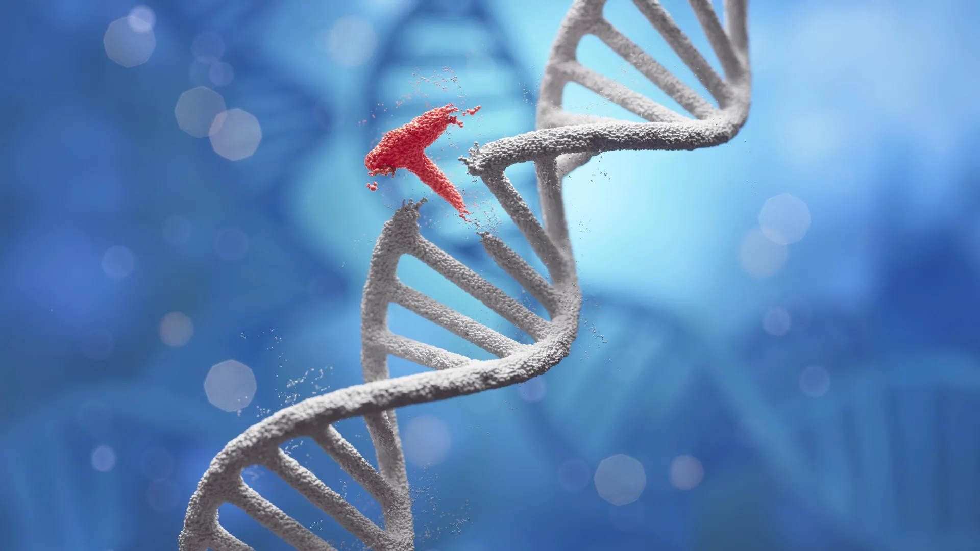
The intricate process of life is encoded within long molecular chains of deoxyribonucleic acid, or DNA, which contain the complete genetic blueprint for every organism. For decades, the field of genetic engineering has sought to harness and modify these instructions, a pursuit that involves meticulously cutting DNA at specific points and seamlessly rejoining these segments with other desired sequences. This fundamental technique underpins a vast array of biotechnological advancements, from enhancing crop resilience and developing novel treatments for inherited diseases to creating sophisticated animal models essential for pharmaceutical research and drug discovery. However, the precise and efficient manipulation of these molecular strands has long presented significant hurdles, particularly when aiming to construct complex or lengthy genetic constructs.
At the heart of successful DNA manipulation lies the ability to create "sticky ends"—short, single-stranded overhangs on DNA fragments that are complementary to other fragments, facilitating their binding. Traditional methods for generating these critical overhangs, and subsequently ligating the DNA pieces, have relied heavily on biological enzymes. Specifically, restriction enzymes are employed to cleave DNA at recognition sites, while DNA ligases, such as T4 DNA ligase, act as molecular glue to seal the newly joined fragments. While foundational, these enzymatic approaches come with inherent limitations. Restriction enzymes are constrained by the specific DNA sequences they can identify and cut, meaning not all desired cleavage points are accessible. Furthermore, the sticky ends they typically produce are often short, usually comprising only a few base pairs. This brevity can significantly diminish the efficiency and stability of the subsequent joining process, posing a considerable challenge for researchers aiming to assemble large or complex DNA molecules with high fidelity. The specificity and short overhangs limit the combinatorial possibilities and the overall speed and accuracy of synthetic DNA construction.
Recognizing these bottlenecks, a collaborative team of scientists from Nagoya University, led by Professor Hiroshi Abe and Assistant Professor Masahito Inagaki, in conjunction with Professor Natsuhisa Oka at Gifu University, embarked on an innovative quest for alternative methodologies. Their focus shifted from biological enzymes to chemical reactions, specifically exploring whether non-enzymatic means could offer a more versatile and efficient route to DNA cleavage and assembly. Their investigation revisited earlier reports from the early 1990s, which demonstrated that silver ions possessed the capacity to cleave 3′-thiol-modified DNA at targeted sites. This historical insight provided a chemical precedent, suggesting a potential pathway to overcome the constraints of enzymatic methods.
Initial experiments leveraging silver ions for DNA cutting proved promising in terms of cleavage effectiveness. However, a critical practical impediment quickly emerged: silver ions exhibited a tendency to bind non-specifically to DNA and induce its precipitation from the solution. This detrimental side effect drastically reduced the recoverable DNA to a mere 14%, rendering the method impractical for real-world applications where high yields of purified DNA fragments are paramount. The challenge, therefore, was not just to cleave DNA, but to do so cleanly and recover the fragments efficiently.
The research team ingeniously hypothesized that replacing free silver ions with silver nanoparticles could resolve the recovery issue. Nanoparticles, being larger and solid, offered the distinct advantage of being physically separable from the reaction mixture, for instance, through centrifugation. This strategy aimed to facilitate the isolation of the desired DNA fragments after cleavage, leaving the silver particles behind. Early tests confirmed the potential of this nanoparticle-based approach. DNA cleavage efficiency reached approximately 50% at 70°C and nearly 100% at 95°C within a two-hour timeframe. While these figures indicated robust cutting, the high temperatures required presented another significant obstacle: prolonged exposure to such heat can denature or damage long DNA molecules, compromising their integrity and utility.
To circumvent the temperature-induced degradation and enhance the overall stability and dispersion of the nanoparticles, the scientists engineered a crucial modification: coating the silver nanoparticles with polyethylene glycol (PEG), a water-soluble polymer. This strategic coating proved transformative. At a more biologically relevant temperature of 37°C, the PEG-modified nanoparticles elevated DNA cleavage efficiency from a modest 36% (without PEG) to an impressive 92% over 31 hours. Further optimization efforts, as highlighted by Assistant Professor Inagaki, the study’s lead author, led to a breakthrough: "In the end, we optimized the conditions to a practical level and, under ambient temperatures, achieved PEG-modified cleaving efficiency above 91% at 50°C within just one to two hours." This significant improvement in efficiency at moderate temperatures marked a pivotal moment, making the nanoparticle method viable for handling delicate DNA molecules.
Beyond enhanced cleavage, the nanoparticle system offered an additional, unexpected advantage: a built-in purification mechanism. The unwanted DNA fragments, after being cleaved, remained adsorbed to the surface of the nanoparticles. In contrast, the precisely cut, desired DNA fragments, complete with their newly formed sticky ends, remained freely suspended in the solution. This differential binding allowed for straightforward separation, dramatically boosting the final DNA recovery rate from the dismal 14% achieved with silver ions to an outstanding 98%. This integrated purification step not only streamlined the experimental workflow but also significantly reduced the loss of valuable genetic material, a common challenge in molecular biology protocols.
Perhaps the most impactful benefit of the silver nanoparticle method lies in its capacity to generate significantly longer sticky ends compared to conventional enzymatic approaches. While restriction enzymes typically produce overhangs of 4 to 6 base pairs, the Japanese team successfully created DNA fragments with 8-base sticky ends using their novel technique. When these longer-overhang fragments were subjected to ligation using T4 DNA ligase, the joining efficiency was approximately double that achieved with traditional methods. The advantages became even more pronounced with extended overhangs; utilizing an 18-base overhang, the researchers recorded a remarkable 44% joining efficiency. This stood in stark contrast to the mere 8% efficiency observed with conventional 4-base overhangs, granting the new nanoparticle-based approach a fivefold improvement in ligation efficacy. The increased length of the sticky ends provides greater specificity and stability to the annealing process, leading to more robust and accurate DNA assembly.
To validate the practical utility of their groundbreaking method in a biological context, the researchers undertook a critical proof-of-concept experiment. They successfully assembled a DNA fragment designed to encode green fluorescent protein (GFP), a widely used reporter gene. Subsequently, this assembled DNA construct was introduced into human HeLa cells. The subsequent observation of successful GFP expression within these cells unequivocally confirmed that the DNA had been accurately constructed and was functionally viable within a living cellular system. This crucial step demonstrated that the nanoparticle-assembled DNA retained its biological activity, paving the way for its application in complex genetic engineering projects.
The implications of this innovative silver nanoparticle technology are far-reaching and hold immense promise for various biotechnological fields. As Assistant Professor Inagaki noted, "We believe this technology will be useful for synthesizing genomic DNA, with many possible applications in areas such as mRNA library establishment for cancer vaccines and gene therapy, as well as the development of artificial protein drugs and genome crops." In gene therapy, the ability to precisely assemble long and complex DNA constructs with high efficiency could accelerate the development of new treatments for genetic disorders by enabling more accurate delivery of therapeutic genes. For cancer vaccines, the creation of diverse and highly specific mRNA libraries could revolutionize immunotherapy strategies. Furthermore, the method could facilitate the production of novel artificial protein drugs with enhanced functionalities and precision. In agricultural biotechnology, it opens new avenues for developing "genome crops" with improved traits, such as disease resistance or increased yield, through sophisticated genetic modifications.
Looking ahead, the research team is focused on expanding the capabilities of their method beyond the assembly of just two DNA fragments at a time. The ultimate goal is to enable the simultaneous joining of multiple fragments, a critical advancement for constructing entire genomes or large synthetic pathways. "We have shown that two DNA fragments can be joined. Now, we need to confirm whether multiple fragments can be joined at the same time—a key step for building genome-scale DNA," explained Inagaki. Achieving this multi-fragment assembly capability would represent a monumental leap forward in synthetic biology, allowing for the rapid and accurate construction of genetic systems of unprecedented complexity. This pioneering work, supported by key agencies such as the Japan Science and Technology Agency (JST) and the Japan Agency for Medical Research and Development (AMED), signifies a transformative step in genetic engineering, offering a robust, efficient, and versatile platform for the precise construction of DNA molecules that could redefine the landscape of biotechnology and medicine.
About the Author