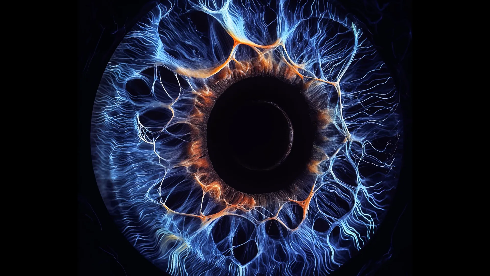
A groundbreaking investigation conducted by researchers at the Yale School of Medicine (YSM) has fundamentally altered long-held assumptions regarding how the human eye processes visual information. Published in the prestigious journal Neuron, the study unveils an intricate web of electrical connections within the retina, suggesting a far more integrated and cooperative system for visual signal processing than previously understood. This paradigm shift in understanding the retina’s internal workings could illuminate mechanisms behind our ability to perceive subtle details or navigate in dimly lit environments, with far-reaching implications for both ophthalmology and broader neuroscience.
For decades, the prevailing scientific consensus maintained that our visual system operates on a principle known as parallel processing. This sophisticated mechanism allows the brain to rapidly deconstruct and interpret the myriad features of any given scene—such as color, motion, contrast, and shape—by dispatching these distinct types of information along separate, specialized neural pathways. The long-standing belief was that these individual channels largely maintained their independence as visual data traversed the retina and proceeded deeper into the brain’s visual cortex. However, the pioneering work from the Yale team challenges this foundational view, revealing that these ostensibly separate channels are, in fact, intimately linked through previously unrecognized electrical connections. This newfound cooperation, according to the research group, appears to play a crucial role in amplifying weak visual signals, preparing them for more robust processing further along the visual cascade.
Dr. Yao Xue, a postdoctoral fellow in YSM’s Department of Ophthalmology and Visual Science and the study’s lead author, articulated the core finding: "Our investigation demonstrated that while various visual channels retain their capacity to deliver specific feature information, they are simultaneously interwoven by an underlying electrical circuitry." This statement encapsulates the revolutionary nature of the discovery, painting a picture of a visual system that is both specialized and profoundly interconnected.
The journey of vision commences at the very back of the eye, within the retina, where specialized photoreceptor cells—rods and cones—are tasked with detecting incoming light. Rods are primarily responsible for vision in low light conditions and peripheral vision, while cones are crucial for color perception and high-acuity vision in bright light. Once activated, these photoreceptors transmit their signals to an intermediate layer of neurons known as bipolar cells. It is at this critical juncture that the raw visual information begins its sorting process, diverging into more than a dozen distinct parallel channels. Each of these channels is dedicated to processing specific visual attributes, ranging from daylight and nighttime vision to color differentiation, contrast sensitivity, and the recognition of shapes.
Traditional understanding largely posited that these bipolar cells communicated primarily via chemical synapses. Neurons typically transmit messages through two primary types of synaptic connections: chemical and electrical. Chemical synapses rely on the release of neurotransmitters—chemical messengers—into a tiny gap between cells (the synaptic cleft), which then bind to receptors on the receiving cell. Electrical synapses, conversely, are direct connections, also known as gap junctions, that allow electrical currents to flow instantaneously and directly from one cell to another. The notion that bipolar cells predominantly utilized chemical communication underscored the assumption of relatively isolated information pathways.
However, when the Yale researchers meticulously examined the synaptic junctions—the microscopic points where bipolar cells transmit information to one another—they uncovered an astonishing revelation. Rather than functioning as isolated conduits, the supposedly independent visual channels were actively exchanging information through a pervasive network of electrical synapses. This unexpected finding was consistent across both mouse and human retinas studied by the team. The implications were profound: when a single bipolar cell was electrically stimulated, the resultant activity was not confined to its immediate pathway but spread significantly, affecting a multitude of other bipolar cell types. Instead of observing neurotransmitter release restricted to a single, narrow channel, the scientists witnessed expansive, diffuse patterns of activity, akin to a "cloud" of communication, unequivocally demonstrating extensive cross-talk between different classes of bipolar cells.
Dr. Z. Jimmy Zhou, the Marvin L. Sears Professor of Ophthalmology and Visual Science and the principal investigator for the study, succinctly summarized this observation: "Upon stimulating a single bipolar cell, we observed that numerous other bipolar cells subsequently released neurotransmitters." This widespread response directly contradicted the independent channel hypothesis.
Further deepening their understanding of this newly discovered network, the research team identified a specific bipolar cell type, designated BC6, which appeared to serve as a central coordinator within this intricate communication web. Signals originating from BC6 cells were observed to propagate across multiple visual pathways in an organized, hierarchical fashion, suggesting a structured rather than random integration of information. Professor Zhou elaborated on this hierarchical arrangement, noting, "Prior to this study, it was largely presumed that the various types of bipolar cells operated with a high degree of autonomy. Our findings, however, point to a ‘driver’ among these cell types—BC6—that actively orchestrates and establishes a hierarchical network."
This dual system—comprising both specialized parallel pathways and overarching electrical communication—offers the retina a sophisticated advantage, leveraging the strengths of both approaches. The specialized channels can efficiently focus on processing distinct visual features, while their interconnectedness enables crucial information sharing, particularly when incoming signals are inherently weak or ambiguous. Dr. Seunghoon Lee, a research scientist in YSM’s Department of Ophthalmology and Visual Science and co-corresponding author of the study, explained the utility of this integration: "If an incoming signal is already very faint and is then further divided across several channels, there is very little residual information for each channel to process effectively. This integration becomes exceptionally valuable for detecting signals of low contrast or those originating from very small objects." Dr. Xue further emphasized the orchestrated nature of this cooperation: "The cells are not collaborating randomly. There is a commander among them—BC6—that guides them in relaying signals to their downstream targets."
Mapping these intricate communication networks posed significant methodological challenges. Bipolar cells are situated deep within the complex architecture of the retina, making them notoriously difficult to access and study in their natural state. Historically, experiments often necessitated dissecting the retina into thin slices to reach these cells, a procedure that inevitably risked disrupting the very neural circuitry researchers sought to examine. To circumvent these limitations and capture the natural dynamics of the system, the Yale team employed a suite of advanced experimental techniques. They utilized sophisticated imaging modalities to precisely monitor how bipolar cells released neurotransmitters and responded to them, while simultaneously stimulating individual cells and recording the responses of their neighboring counterparts.
A pivotal innovation in this study was the successful application of a dual patch clamp technique on fully intact mouse retinas. This highly demanding electrophysiological method involves precisely positioning two microscopic electrodes onto individual cells to both stimulate and record electrical activity, all while maintaining the retina’s complete structural and functional integrity. Professor Zhou lauded this technical achievement, stating, "No other laboratory globally has consistently managed to execute these types of recordings with such systematic rigor. It represents an exceptional feat of Dr. Yao Xue’s doctoral research, combining an inventive methodological approach with unparalleled electrophysiological expertise." The team subsequently replicated these experiments using intact human retinas, obtained through the Department of Pathology’s Legacy Tissue Donation Program. These human retina experiments mark the first instance of such detailed, intact-tissue investigations, significantly bolstering the translational relevance of the findings.
The ramifications of this discovery extend far beyond the realm of vision science. Given that the retina is an integral, accessible extension of the central nervous system (CNS), these findings offer a unique window into the fundamental organizational principles of neural networks. Understanding how retinal circuits efficiently process and integrate information could provide invaluable new insights into the operational mechanisms of other complex neural structures throughout the brain. This could potentially inform our understanding of various neurological disorders that involve synaptic dysfunction or abnormal neural connectivity.
Moreover, the research holds direct relevance for improving our comprehension and treatment of numerous diseases that progressively damage the retina, including age-related macular degeneration, glaucoma, and inherited forms of congenital night blindness. By elucidating the precise mechanisms of signal integration and amplification, scientists may uncover novel therapeutic targets for preserving or restoring visual function in affected individuals.
Finally, the researchers underscored the profound value of curiosity-driven scientific inquiry. Rather than commencing with a predefined hypothesis, this research journey was propelled by fundamental questions, ultimately uncovering a previously unknown processing mechanism that reshapes our entire understanding of visual perception. Dr. Lee emphasized this philosophical aspect: "Our experiments did not originate from a specific hypothesis but instead revealed a fundamental processing mechanism within the visual system. It serves as an important reminder of the essential role that curiosity-driven research plays in driving true scientific discovery." This landmark study from Yale stands as a testament to the fact that even in seemingly well-understood biological systems, profound and unexpected secrets continue to await discovery, offering new pathways for knowledge and therapeutic innovation.
About the Author