
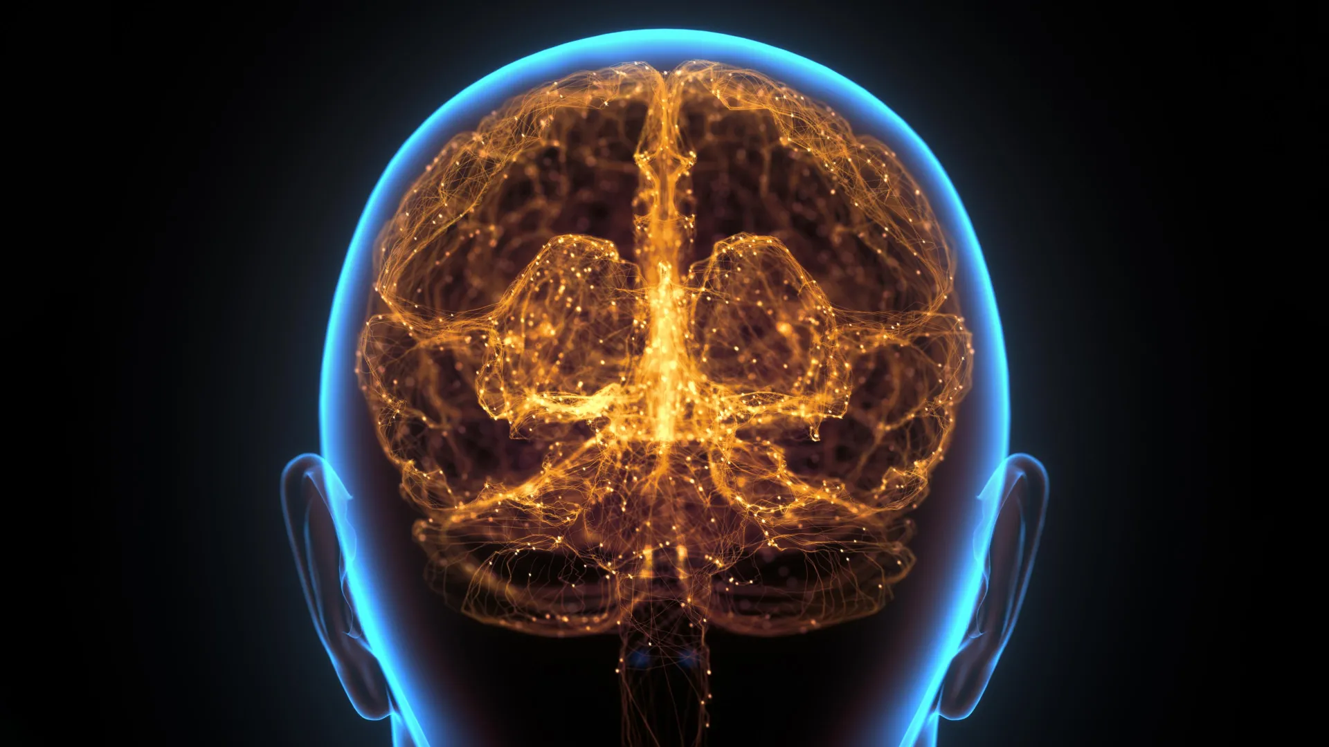
For decades, the prevailing scientific consensus posited a remarkable degree of immunological seclusion for the brain, shielding it from the body’s broader defense mechanisms. However, a groundbreaking investigation conducted by scientists at Washington University School of Medicine in St. Louis has dramatically upended this long-held view, revealing a previously unrecognized and highly specialized immune infrastructure nestled within the bone marrow of the skull. These newly identified structures, exhibiting characteristics akin to lymph nodes, appear to serve as vigilant sentinels, capable of mounting a swift and targeted response to the emergence of cancerous growths within the brain, predating even the activation of more distant lymphatic glands.
The research team’s meticulous work, detailed in a seminal publication in the prestigious journal Nature, not only identified these intricate immune congregations in laboratory mice but also uncovered compelling evidence of their existence in human subjects. This parallel observation strongly suggests that the localized immune surveillance system identified in rodents is not an anomaly of animal physiology but potentially a conserved mechanism across species, including our own. The implications of this discovery are profound, fundamentally reshaping our understanding of how the central nervous system interacts with the immune system and opening novel therapeutic avenues for a range of debilitating neurological conditions.
The scientific journey leading to this revelation has been a progressive dismantling of established dogma. Earlier foundational research from the laboratory of senior author Jonathan Kipnis, PhD, a distinguished professor and investigator at WashU Medicine, began to erode the notion of the brain’s complete immune isolation. A pivotal earlier finding by Kipnis’s team was the identification of lymphatic vessels embedded within the dura mater, the tough, outermost protective membrane enveloping the brain, situated directly beneath the skull. This discovery provided the first tangible evidence of direct vascular connections facilitating communication between the brain’s protective layers and the peripheral immune system.
Building upon this foundational work, subsequent investigations uncovered an even more intimate anatomical link: microscopic channels that traverse the cranial bone, bridging the dura and the brain tissue itself. These minuscule conduits act as direct highways, enabling the bidirectional movement of immune cells and cellular debris between the central nervous system and the adjacent skull bone marrow. It is through these pathways that the newly discovered immune hubs are thought to receive vital information about events occurring within the brain.
In the current study, researchers meticulously tracked the movement of specific proteins originating from the brain, observing their passage through these cranial channels and their subsequent arrival within the skull bone marrow. It was within this osseous environment that they encountered highly organized immune structures that bear a striking resemblance to the architecture and function of lymph nodes, critical components of the body’s adaptive immune system. Lymph nodes are renowned for their role as central command centers for immune responses, facilitating crucial interactions between various immune cell types. Within these nodes, specialized cells, such as T follicular helper cells, collaborate with B cells, which are responsible for generating antibodies – the molecular soldiers that target and neutralize pathogens and abnormal cells. The presence of such sophisticated immunological organization within healthy bone marrow was an entirely unexpected and groundbreaking observation.
Jang Hyun Park, PhD, the study’s lead author and a postdoctoral research fellow whose work has culminated in this significant finding, expressed the team’s astonishment. "We have never witnessed such structures within healthy bone marrow prior to this research," Dr. Park stated, noting that his upcoming tenure at the Korea Advanced Institute of Science and Technology will be informed by these transformative insights. He further elaborated, "This is an exhilarating discovery that underscores the intricate needs of a complex organ like the brain, which appears to necessitate its own dedicated immune structures for robust defense."
The researchers then turned their attention to assessing the functional relevance of these cranial immune centers, specifically their role in combating disease. Employing a meticulously designed mouse model engineered to mimic glioblastoma, an exceptionally aggressive form of brain cancer, the team deliberately disrupted the function of these skull-based immune hubs. The results were stark: tumors exhibited accelerated growth in mice where these local immune defenses were compromised, compared to their counterparts with intact cranial immune structures. Furthermore, the mice with impaired skull immune surveillance displayed a significantly reduced survival rate, providing compelling empirical evidence that these localized immune outposts are not merely passive bystanders but actively contribute to the body’s fight against brain malignancies.
Intrigued by the potential for therapeutic intervention, the scientists explored whether the immune response originating from these cranial hubs could be deliberately amplified. To this end, they developed an innovative therapeutic strategy designed to enhance antibody production within the skull bone marrow. This approach involved combining three potent immune-stimulating proteins, formulating them into a gel, and applying this agent directly to the scalp, thereby facilitating its proximity to the cranial bone marrow.
The application of this targeted treatment elicited a robust and immediate surge of immune activity directed against the experimental tumors. Crucially, the researchers observed that this heightened immune response manifested first within the specialized immune hubs located inside the skull bone marrow, only subsequently appearing in more distant lymph nodes situated outside the cranial vault. This temporal and spatial pattern of activation underscores the primary role of the skull’s immune structures as the initial responders. The treated mice demonstrated a superior capacity to reject tumors and exhibited a marked improvement in survival duration when contrasted with the control group, highlighting the efficacy of this localized immune modulation strategy.
The implications of this landmark discovery extend far beyond the realm of brain cancer, promising to revolutionize the approach to a broad spectrum of neurological disorders. Given the direct anatomical proximity of these immune hubs to the brain, researchers posit that they could offer a novel and highly targeted pathway for influencing immune activity within the central nervous system. This localized approach could potentially modulate immune responses in conditions such as Alzheimer’s disease, Parkinson’s disease, schizophrenia, and even complex conditions like long COVID, all of which are believed to involve an inflammatory or immune component, without triggering widespread systemic immune alterations that often accompany conventional treatments.
"This finding fundamentally alters our current paradigm in neuroimmunology," stated Professor Kipnis. "Understanding that the brain relies on these first responders located within the surrounding cranial bone for its defense has the potential to profoundly reshape how we conceive of and develop therapeutic interventions for a multitude of neurological conditions. The prospect of directly accessing these immune hubs through the skull, bypassing the need for systemic administration and thereby minimizing peripheral side effects, represents a truly transformative advance in the field." This discovery heralds a new era in our understanding of brain-immune interactions and offers tangible hope for more effective and less invasive treatments for some of humanity’s most challenging diseases.
About the Author












