The human capacity to acquire and retain new abilities is fundamental to adaptation and growth, yet the mechanisms governing how transient practice transforms into enduring expertise remain a profound area of scientific inquiry. While individual aptitude and diligent effort are widely recognized as contributors to mastery, emerging neuroscientific research increasingly suggests that the brain’s physiological state, particularly its interaction with the body, plays a critical, yet often overlooked, role in solidifying learned behaviors. A groundbreaking study from Tohoku University in Japan now sheds light on this intricate connection, demonstrating that stimulation of the vagus nerve following training sessions can significantly bolster long-term motor learning, specifically by influencing rhythmic changes in the brain’s vascular environment. This pivotal research, published in the journal iScience on August 25, 2026, posits a fresh perspective on how the body’s internal signals contribute to the brain’s capacity for lasting change.
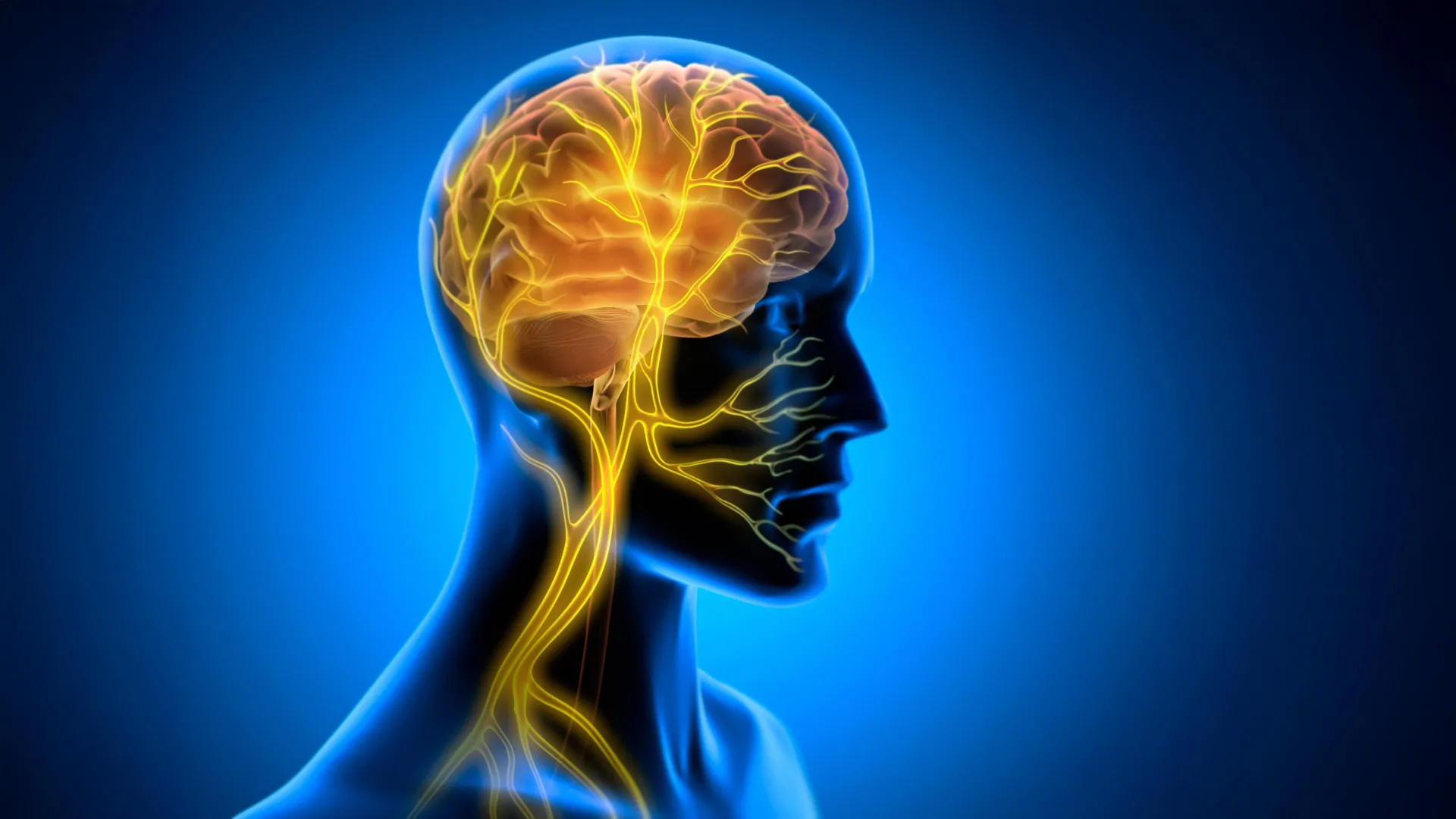
For decades, the vagus nerve has captivated researchers due to its extensive reach and multifaceted influence across numerous bodily systems. As the longest cranial nerve, it forms a crucial, bidirectional communication highway, transmitting sensory information from the visceral organs—including the heart, lungs, and gastrointestinal tract—to the brain, while simultaneously relaying motor and autonomic commands from the central nervous system back to these organs. This anatomical and functional centrality has made the vagus nerve a prime target for therapeutic interventions, particularly through vagus nerve stimulation (VNS). Clinically approved for a range of conditions, including epilepsy and severe depression, VNS typically involves implanting a device that delivers mild electrical pulses to the nerve, a process known as neuromodulation, which is believed to exert its effects primarily by altering neurotransmitter systems within the brain.
However, the team at Tohoku University, specializing in super network brain physiology, hypothesized that the vagus nerve’s role in learning might extend beyond simple neurotransmitter modulation. Their investigation delved into the possibility of a different, more dynamic mechanism: the influence of VNS on the brain’s vascular system. This represents a significant conceptual leap, suggesting that the rhythmic ebb and flow of blood within cerebral vessels could be a critical, previously unrecognized factor in how the brain consolidates new motor skills.
To rigorously test this hypothesis, the scientists developed a sophisticated experimental paradigm using mice as their model subjects. They meticulously designed a miniature cuff electrode, carefully positioned to remain attached to the left cervical vagus nerve. This allowed for precise and controlled delivery of electrical stimulation. The learning task chosen for the study was the horizontal optokinetic response (HOKR), an eye movement task that relies heavily on cerebellar function. The HOKR task requires mice to refine their ability to follow moving visual stripes, mirroring the involuntary eye adjustments humans make when tracking a moving object, such as a passing train from a stationary platform. This cerebellar-dependent task served as an ideal model for assessing motor learning and plasticity.
Crucially, the researchers deviated from conventional VNS application by administering the stimulation after each training session, rather than concurrently with the learning task itself. This deliberate timing was predicated on the understanding that while initial skill acquisition occurs during practice, the transformation of this nascent knowledge into durable, long-term memory—a process known as memory consolidation—primarily takes place during subsequent rest periods. The immediate behavioral observations confirmed this strategic approach: VNS did not confer any immediate performance enhancement during the training sessions. The true impact of the stimulation manifested later, in the days following the initial learning phase.
Remarkably, mice that had received VNS after their training consistently exhibited superior long-term learning compared to their unstimulated counterparts. This delayed but robust benefit strongly implied that the vagus nerve stimulation was not directly influencing the act of learning but rather facilitating the consolidation processes that occur in the brain post-training. Professor Ko Matsui, a senior author on the study, articulated this insight, emphasizing that "the pivotal aspect was that VNS was administered exclusively after training." He further elaborated, "Our discoveries intimate that vagus nerve stimulation might unveil a latent window of opportunity for augmented learning by rendering the neural environment more receptive to enduring modifications." This statement underscores the notion that VNS could be priming the brain for optimal memory encoding and retention during critical post-practice periods.
To unravel the underlying physiological mechanisms responsible for this enhanced learning, the research team employed fiber photometry to monitor real-time changes in blood volume activity within the cerebellar flocculus, an area of the brain specifically implicated in HOKR learning. Their detailed measurements revealed a distinct, two-phase vascular response triggered by a single application of VNS. Initially, a brief, localized decrease in blood volume was observed, which was then followed by a delayed but significant increase. When VNS was administered repeatedly, these vascular dynamics evolved into rhythmic oscillations in blood volume within the cerebellar region.
Even more compelling was the direct correlation observed between these vascular rhythms and learning outcomes. Mice exhibiting more pronounced and robust blood volume oscillations generally demonstrated superior learning by the fifth day of the experiment. This finding strongly suggests a direct link between the brain’s vascular environment and the long-term efficacy of the vagus nerve stimulation, pointing to a novel mechanism through which VNS might exert its cognitive benefits. The hypothesis emerging from this data is that these rhythmic changes in cerebral blood flow could be optimizing the metabolic support to neurons, facilitating waste clearance, or even acting as a signaling mechanism that promotes synaptic plasticity—the strengthening or weakening of connections between neurons that underlies learning and memory.
Junyu Chen, the lead author of the study, reflected on the broader implications of these findings: "Our brains may be more profoundly influenced by the body than we currently appreciate." He further posited, "By finely tuning the brain’s metabolic landscape, which includes these rhythmic vascular movements, we may eventually unlock cognitive capacities that might otherwise remain dormant." This perspective challenges the traditional view of the brain as an isolated processing unit, instead presenting it as an organ deeply intertwined with and responsive to the physiological states of the entire body. The research posits that the vagus nerve serves as a critical conduit for this integration, translating bodily signals into neurovascular changes that directly impact cognitive function.
Looking ahead, the Tohoku University team plans to expand on these foundational discoveries. Future investigations will concentrate on refining the stimulation protocols, aiming to determine the most effective parameters for VNS to maximize its learning-enhancing effects. A key objective will be to delineate with greater precision the exact molecular and cellular pathways through which this brain-body communication supports long-term plasticity. This will involve exploring how vascular oscillations translate into changes at the synaptic level and how these processes contribute to the structural and functional modifications in neural networks that underpin lasting skill acquisition.
The potential implications of this research are far-reaching. By gaining a more comprehensive understanding of this intricate, two-way communication pathway between the brain and the body, scientists hope to not only decipher the fundamental mechanisms of how learning becomes permanent but also to develop innovative strategies for its enhancement. This could pave the way for novel therapeutic approaches in areas such as neurorehabilitation, aiding patients recovering from stroke or traumatic brain injury to regain motor function more effectively. It could also have profound applications in educational settings, optimizing learning strategies, or even in fields requiring high-level skill acquisition, such as athletic training or complex professional development. The work by Tohoku University represents a significant stride in bridging the gap between peripheral physiology and central cognitive function, offering exciting new avenues for unlocking the full potential of human learning and memory.
About the Author