
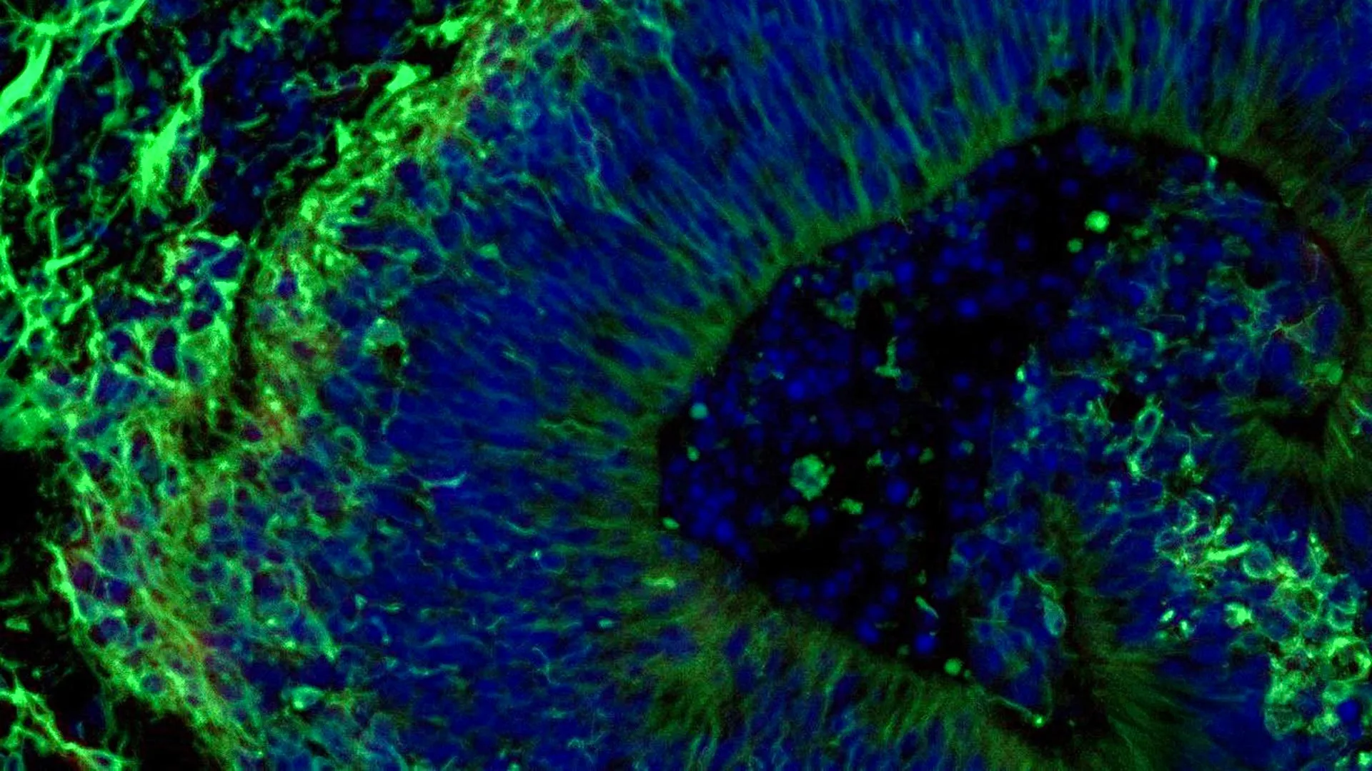
Scientists at Johns Hopkins Medicine have presented compelling new findings suggesting that intricate, lab-cultivated brain tissue models, derived from individuals diagnosed with Alzheimer’s disease, hold significant potential for forecasting patient responses to pharmacological interventions aimed at alleviating associated neuropsychiatric manifestations. This groundbreaking research contributes to a growing body of evidence advocating for the utility of these miniature brain constructs, known as organoids, in facilitating the development and selection of highly personalized therapeutic strategies for distinct patient cohorts affected by Alzheimer’s, the predominant form of dementia impacting over seven million Americans. Beyond their predictive capabilities, the investigation also illuminated the release of minuscule cellular packages, termed extracellular vesicles, from these organoids, which may serve as novel diagnostic markers for the disease and indicators of its progression. The study, partially supported by the National Institutes of Health, was formally documented in Alzheimer’s & Dementia: The Journal of the Alzheimer’s Association.
The implications of this research are profound, pointing towards a future where clinical decision-making for Alzheimer’s patients could be significantly enhanced by personalized laboratory assessments. Dr. Vasiliki Machairaki, the lead investigator and an associate professor of genetic medicine at the Johns Hopkins University School of Medicine, articulated the study’s central hypothesis: "Our work suggests that large-scale, patient-derived brain organoids, along with the vesicles they release, can provide a sophisticated platform for staging Alzheimer’s disease, delving into its underlying pathological mechanisms, and crucially, evaluating how specific patient subgroups might react to various therapeutic agents." Currently, Alzheimer’s remains an incurable condition, though treatments like selective serotonin reuptake inhibitors (SSRIs) are frequently employed to manage challenging neuropsychiatric symptoms such as anxiety, depression, and agitation, which are experienced by nearly all individuals with the disease. However, the efficacy of these medications exhibits considerable variability among patients, a phenomenon this research aims to unravel.
The investigative team focused their efforts on developing and analyzing hindbrain organoids, miniature representations of the brain region responsible for regulating vital autonomic functions like respiration, sleep cycles, and heart rate. The primary objective was to ascertain whether these models could reveal molecular signatures that predict whether escitalopram oxalate, a specific SSRI, might effectively mitigate the symptomology associated with Alzheimer’s disease. To achieve this, researchers commenced by obtaining blood samples, with informed consent, from individuals diagnosed with Alzheimer’s disease through the NIH-funded Johns Hopkins Alzheimer’s Disease Research Center. These collected blood cells were then meticulously reprogrammed back to an induced pluripotent stem cell (iPSC) state, a versatile progenitor cell type capable of differentiating into virtually any cell within the human body.
Utilizing iPSCs sourced from both Alzheimer’s patients and healthy control participants, the researchers meticulously cultivated hindbrain organoids. These organoids were engineered to contain specialized neural cells, or neurons, that are capable of synthesizing serotonin, a critical neurotransmitter. Through precise biochemical guidance, these stem cells were encouraged to self-organize into small, approximately pea-sized, three-dimensional clusters of brain tissue that closely mimic the structural characteristics of the hindbrain. The study’s impressive scale involved hundreds of these organoids, each meticulously representing an individual patient or healthy subject, positioning it as one of the most extensive brain organoid studies conducted to date within the Alzheimer’s research landscape.
Upon maturation, the patient-derived organoids exhibited several key biological hallmarks characteristic of Alzheimer’s disease at a molecular level. When compared to organoids developed from healthy donors, those originating from Alzheimer’s patients displayed discernible alterations in proteins integral to neuronal communication, inflammatory processes, and pathways known to be implicated in the disease’s pathogenesis. Subsequently, these organoids were exposed to escitalopram oxalate, a widely prescribed antidepressant medication. The observations revealed that in a subset of organoids derived from Alzheimer’s patients, the administration of the medication led to an augmentation of proteins associated with serotonin signaling and intercellular communication within the brain. These are precisely the biochemical pathways that antidepressant therapies are designed to modulate. Conversely, other organoids demonstrated minimal to no discernible molecular response to the treatment. Dr. Machairaki commented on this variability, stating, "We employed these organoids to model how tissue from certain patients might respond to a commonly prescribed SSRI. On a broad scale, our model holds the promise of eventually identifying patient subgroups, based on their unique molecular underpinnings, who are more likely to benefit from specific drugs, thereby paving the way for the development of highly precise and targeted treatments in the long term."
The research further explored the potential utility of extracellular vesicles, secreted by the organoids, as diagnostic biomarkers for Alzheimer’s disease or as indicators of therapeutic response. Scientists meticulously analyzed the protein composition within extracellular vesicles released by both patient-derived and healthy control organoids, both prior to and following escitalopram treatment. These vesicles were found to contain proteins crucial for fundamental brain functions, including synaptic transmission, memory formation, and neurotransmitter release. Organoids cultivated from individuals with Alzheimer’s disease exhibited significant deviations in several proteins associated with the disease state, with notably lower levels of RAB3A, NSF, and ATCAY observed. These proteins play pivotal roles in the normal functioning of neuronal signaling. Post-escitalopram administration, certain protein levels experienced an increase in specific samples, with the most pronounced changes observed in proteins linked to serotonin signaling and synaptic pathways targeted by antidepressants. The differential responses observed across organoids – some showing robust molecular engagement with the drug and others displaying negligible changes – strongly suggest that extracellular vesicles derived from brain organoids could ultimately serve as a tool for predicting which patients are most likely to achieve therapeutic benefits from a particular medication.
Looking ahead, Dr. Machairaki outlined plans for the development of more sophisticated organoid models. These advanced constructs are intended to incorporate immune cells and vascular-like networks that closely recapitulate the intricate architecture of blood vessels found in living human brain tissue. The integration of these components is anticipated to enhance the fidelity of these laboratory models, making them even more representative of actual human brain environments. With continued research, the aspiration is that extracellular vesicles could eventually function as a non-invasive diagnostic tool, akin to a liquid biopsy, potentially aiding in the early detection of Alzheimer’s disease, the accurate staging of its progression, and the precise identification of individual disease subtypes. Dr. Machairaki underscored that the present study represents a foundational step towards realizing this ambitious objective. The collaborative effort involved a multidisciplinary team, including researchers from Johns Hopkins University, Tymora Analytical Operations, and the University of Rochester School of Medicine and Dentistry, with substantial funding provided by various grants from the National Institutes of Health, the Paul G. Allen Frontiers Foundation, and the Richman Family Precision Medicine Center of Excellence in Alzheimer’s Disease at Johns Hopkins University. No conflicts of interest were declared by any of the authors under the established policies of Johns Hopkins University.
About the Author












