Unveiling a Novel Immune System Dysfunction Linked to Dental Implant Degradation.
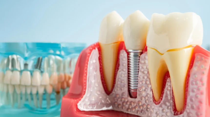
For a significant portion of the population, dental implants represent a sophisticated solution for restoring oral function and aesthetics following tooth loss, offering a more permanent and stable alternative to conventional dentures. However, a notable percentage of individuals, estimated between 10% and 20% of those who receive implants, can develop a severe complication known as peri-implantitis. This inflammatory condition not only compromises the integrity of the surrounding gum tissue but also leads to a progressive destruction of the jawbone that anchors the implant, posing a considerable challenge to restorative dentistry.
A recent groundbreaking study, published in the esteemed journal PNAS Nexus, has illuminated a previously poorly understood mechanism underlying the recalcitrance of peri-implantitis to standard antibiotic treatments. Researchers affiliated with the Rutgers School of Dental Medicine have identified a critical pathway through which microscopic titanium particles, shed from degrading dental implants, profoundly disrupt the body’s natural defense mechanisms. This disruption leads to a chronic inflammatory state that ultimately undermines the bone structure essential for implant stability.
The investigative team employed a multi-pronged approach, integrating the analysis of human tissue samples obtained from patients, in vitro experiments with human immune cells, and sophisticated studies utilizing genetically modified mouse models. Their meticulous work pinpointed a specific calcium channel within macrophages, the versatile immune cells tasked with engulfing and neutralizing pathogens. The presence of titanium particles was found to activate this channel, effectively trapping the macrophages in a hyper-inflammatory cycle. Crucially, when this particular calcium channel was experimentally disabled in the mouse subjects, the development of the destructive inflammatory disease was notably prevented.
This significant scientific breakthrough offers the first concrete molecular target for therapeutic intervention in a condition that affects a substantial segment of implant recipients and incurs considerable healthcare expenditures globally, with estimates placing the annual cost to healthcare systems in excess of one billion dollars. Dr. Georgios Kotsakis, the senior author of the study and assistant dean for clinical research at Rutgers School of Dental Medicine, emphasized the paradigm shift this discovery represents. "For the first time, we are able to articulate why the antibiotic regimens that prove effective in managing infections around natural teeth fail to yield similar results when applied to the tissues surrounding dental implants," he stated. "With this foundational understanding of the root cause, we can now embark on the development of targeted therapeutic strategies."
A persistent enigma in the field of peri-implantitis has been its striking resemblance to periodontitis, the more common gum disease affecting natural teeth. Both conditions are initiated by similar oral bacteria and manifest with inflammation and bone loss. However, their responses to treatment diverge dramatically. While antibiotics, coupled with meticulous professional cleaning, can frequently resolve periodontitis, the same interventions achieve success in fewer than half of peri-implantitis cases. Even with aggressive treatment, the underlying bone deterioration can often continue unabated.
For the past two decades, the scientific community’s focus for understanding peri-implantitis had largely been concentrated on the microbial agents involved. In contrast, the research group led by Dr. Kotsakis chose to explore an alternative hypothesis: the potential role of the implant material itself in fostering the disease process. Their investigations revealed that the bacterial biofilms that inevitably form on the surfaces of dental implants can generate acidic microenvironments. These acidic conditions, over time, contribute to the slow corrosion of the titanium alloy, the material commonly used in dental implants. This corrosive process can liberate a vast quantity of microscopic metal particles, each significantly smaller than a human red blood cell, into the adjacent soft tissues and bone.
Compounding this issue, titanium particles can also be inadvertently released during routine dental hygiene procedures. This risk is particularly elevated when instruments designed for cleaning natural teeth are employed on implants. Such abrasive tools can inadvertently abrade and damage the implant surface, dislodging more of these nano-sized metal fragments and exacerbating the problem.
The intricate process by which these liberated titanium fragments exert their detrimental influence on the immune system has now been elucidated. Once dispersed within the gingival tissues, the minute titanium particles possess an affinity for lipopolysaccharide (LPS), a potent endotoxin produced by certain bacteria commonly found in the oral cavity. This interaction effectively coats the titanium fragments with bacterial components. The immune system, encountering these particles, perceives them as unusually large and indigestible foreign entities, akin to significantly overgrown bacteria that cannot be efficiently processed.
Macrophages, the frontline phagocytic cells of the immune system, are then recruited to engulf these titanium-LPS complexes. However, their inability to degrade the inert titanium material leads to a prolonged and unproductive state of heightened inflammation. In this chronic inflammatory state, macrophages release excessive amounts of pro-inflammatory signaling molecules, most notably interleukin-1 beta (IL-1β). This cytokine is well-known for its involvement in various inflammatory conditions, including autoimmune disorders like rheumatoid arthritis and neurodegenerative diseases such as Alzheimer’s disease.
The sustained inflammatory cascade triggered by the titanium particles gradually leads to the enzymatic breakdown of the bone matrix, compromising the osseointegration—the crucial biological process by which bone fuses with the implant. Simultaneously, the macrophages become significantly less effective in their primary role of combating the actual bacterial pathogens that initiated the infection. Laboratory observations starkly illustrated this functional impairment: macrophages exposed to titanium particles demonstrated less than half the capacity to engulf bacteria compared to their counterparts that had not been exposed to the metallic debris. Dr. Kotsakis eloquently summarized this phenomenon: "These particles act as molecular magnets, attracting bacterial toxins and consequently hijacking the immune system, thereby incapacitating its ability to eradicate the invading bacteria. The result is a perfect storm that renders antibiotic therapies ineffective."
Following the revelation of how titanium particles disrupt immune surveillance and function, the researchers delved deeper into the molecular underpinnings of this destructive inflammatory response. Their investigation traced the key signaling pathway to TRPC1, a specific type of calcium channel embedded within the cell membranes of macrophages. This channel plays a pivotal role in mediating the inflammatory cascade initiated by the combined presence of titanium particles and bacterial toxins.
To rigorously test the significance of TRPC1 in this pathological process, the research team utilized genetically engineered mice engineered to lack this particular calcium channel. These TRPC1-deficient mice were then subjected to the same experimental conditions involving titanium particles and bacteria. The results were remarkable: the immune systems of these mice exhibited a far more robust and controlled response. They developed significantly smaller inflammatory lesions, produced substantially lower levels of inflammatory cytokines, and retained their capacity to effectively clear bacterial populations. The experimental disabling of the TRPC1 channel effectively abrogated the destructive inflammatory disease that was readily observed in the presence of a functional TRPC1 channel.
These findings collectively point towards a promising new therapeutic avenue for managing peri-implantitis, one that focuses on modulating the dysregulated immune response rather than relying solely on conventional antibiotic approaches. Supported by funding from the National Institutes of Health, Dr. Kotsakis and his team are currently engaged in evaluating various pharmacological agents designed to target this specific molecular pathway within human cell cultures.
While this research opens exciting possibilities for future treatments, it also serves to underscore the critical importance of established preventive measures for individuals who have received dental implants. Consistent and professional dental cleanings remain the most potent known defense against the onset and progression of peri-implantitis. However, the specific techniques and instruments employed during these appointments are of paramount importance. Until relatively recently, many dental practitioners routinely utilized metal scalers, identical to those used for natural teeth, to debride implants. However, subsequent research, including that conducted by the Rutgers team, has conclusively demonstrated that these abrasive tools can damage and corrode titanium surfaces, potentially leading to the release of additional particles and accelerating the disease process. In contemporary dental practice, the widespread adoption of non-abrasive cleaning methods has become the standard of care, thereby enhancing the protection of implant surfaces and mitigating the risk of complications. The comprehensive understanding provided by this latest research, detailing how corroded titanium can interfere with host immune defenses, offers a novel explanation for the persistent challenges in treating certain dental implant infections and simultaneously charts a promising course for the development of more effective therapeutic interventions.

Related News