Congenital heart disease (CHD) represents a significant global health challenge, affecting approximately two out of every 100 live births worldwide. This makes it one of the most frequently diagnosed birth defects, leading to structural abnormalities in the heart that originate during the intricate process of embryonic development. Despite its prevalence and profound impact on millions of newborns and their families each year – with an estimated 2.3 to 2.5 million infants affected annually and around 16 million individuals living with the condition as of 2023 – the precise molecular and cellular mechanisms underlying these cardiac malformations have largely remained elusive. The complexity of early human development, particularly the formation of vital organs like the heart, presents a formidable frontier for scientific investigation.
However, recent pioneering research conducted by scientists at the University of Copenhagen has shed new light on this enduring mystery. Their investigations have uncovered a previously unrecognized cellular communication network that appears to play a pivotal role in ensuring the proper formation of the heart during its embryonic stages. This groundbreaking discovery offers a critical piece to the intricate puzzle of cardiac development and promises to reshape our understanding of why certain congenital heart defects manifest.
Professor Lars Allan Larsen, a leading authority on congenital heart disease at the Department of Cellular and Molecular Medicine, emphasized the significance of these findings. "We have identified a novel communication system situated on the exterior of cells that is absolutely essential for the heart’s correct architectural formation within the developing embryo. This revelation fundamentally alters our perspective on the origins of some congenital heart defects. It’s akin to pinpointing a crucial gear in an exceptionally complex biological machinery."
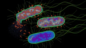
The newly identified mechanism operates within the primary cilium, a remarkable and ubiquitous cellular structure. Often described as a microscopic, hair-like projection, the primary cilium extends from the surface of nearly every cell type within the human body. Far from being a mere decorative appendage, these specialized organelles function as sophisticated cellular antennae, enabling cells to perceive and interpret a diverse array of chemical signals emanating from their microenvironment. These signals are not trivial; they are critical inputs that can dictate fundamental cellular decisions, including whether a cell should proliferate, migrate, differentiate into a specific tissue type, or undergo programmed cell death. Their widespread presence and sensory capabilities underscore their importance across various physiological processes, particularly during the rapid and highly coordinated events of embryonic organogenesis.
Within the intricate architecture of this cellular sensory organelle, the researchers pinpointed a crucial signaling nexus. They discovered that three specific proteins – designated TAK1, TAB2, and PKA-Cα – collaborate synergistically to form a regulatory hub inside the primary cilium. The synchronized activity of these proteins appears to be indispensable for the normal progression of cardiac development. Disruptions to this delicate balance can have profound consequences for the nascent heart.
Professor Søren Tvorup Christensen, a cell biologist from the Department of Biology, elucidated the functional role of this protein trio. "These proteins essentially serve as molecular directives, guiding stem cells on the precise timing and manner of their differentiation into specialized heart muscle cells. When genetic alterations compromise this intricate communication pathway, it can result in ‘ciliary defects,’ which subsequently may lead to the manifestation of congenital heart anomalies." This explanation highlights how even subtle genetic variations impacting these proteins can derail the meticulously choreographed process of heart formation.
To rigorously investigate this novel cellular system, the research team employed a multi-pronged approach, integrating diverse methodologies. Their investigation commenced with a comprehensive analysis of genetic data derived from several thousand individuals diagnosed with congenital heart defects. The scientists meticulously searched for rare genetic mutations, comparing their prevalence in affected patients against healthy control subjects. Genetic variants that exhibited a statistically significant higher frequency among patients were flagged as strong candidates for contributing to the condition. This initial genetic screening provided crucial leads, directing subsequent experimental inquiries.
Building upon these genetic insights, the team then moved to functional validation using advanced genetic engineering techniques. They meticulously recreated the identified genetic mutations in zebrafish, a widely recognized and powerful vertebrate model organism for developmental biology studies due to its rapid external development and genetic tractability. By observing the impact of these induced mutations, the researchers were able to assess how these genetic changes influenced heart formation in a living organism. The results compellingly demonstrated that alterations in these specific genes could indeed disrupt normal cardiac development and compromise heart function within the zebrafish models.
Further strengthening their findings, the researchers also conducted extensive laboratory experiments utilizing various types of cells, including human cell lines and mouse stem cells. These in vitro studies provided a granular view of the signaling system, enabling the scientists to dissect its molecular intricacies and precisely determine the cascade of events that transpires when its communication pathways are perturbed. The convergence of evidence from patient genetics, in vivo zebrafish experiments, and in vitro cellular models consistently pointed towards the primary cilium and its internal signaling hub as a critical determinant in the pathogenesis of congenital heart defects.
Professor Larsen expressed strong confidence in the findings, stating, "We approached this mechanism from numerous perspectives and utilized a wide array of experimental methods. The consistency of our observations across all these different angles, critically aligning with what we observe in human patients, gives us considerable assurance that this mechanism is indeed active and relevant in human development." This emphasis on translational relevance underscores the potential impact of their discovery.
The implications of this research extend far beyond the confines of cardiac development. The team also uncovered compelling evidence suggesting that this newly identified ciliary mechanism may influence the proper formation of other vital organs throughout the body. The rare mutations initially scrutinized in this study were predominantly identified in individuals diagnosed with what is known as syndromic congenital heart disease. In these complex cases, a heart defect is not an isolated anomaly but rather forms part of a broader genetic syndrome, which often manifests with additional abnormalities affecting other organ systems.
Through further experiments in zebrafish and detailed investigations of primary cilia in various tissues, the researchers gathered evidence indicating that the same fundamental cellular mechanism could contribute to the development of multiple organs simultaneously. This provides a crucial explanatory framework for previously puzzling clinical observations.
Professor Christensen elaborated on this broader impact: "When the ciliary mechanism falters, it typically impairs the development of several other organs in conjunction with the heart. This observation provides a unifying explanation for why some patients with congenital heart disease also present with defects and associated conditions affecting the brain, kidneys, and skeletal system. The mechanism offers a cohesive understanding for a constellation of diseases that we have historically struggled to fully comprehend."
Given that dysfunctions involving the primary cilium are already recognized as causative factors in numerous rare genetic disorders, collectively known as ciliopathies, the researchers anticipate that this new discovery could significantly enhance the scientific community’s understanding of a much wider spectrum of human diseases. Ciliopathies are a diverse group of genetic conditions characterized by a broad range of symptoms affecting various organs, precisely because primary cilia are so fundamental to cellular function and development across the body.
"Many rare genetic conditions are attributed to genetic alterations that impair ciliary function, yet the precise underlying molecular mechanisms have often remained obscure," Professor Larsen noted. "This new knowledge could eventually facilitate earlier identification of affected patients and pave the way for the development of more targeted and effective therapeutic interventions." Such advancements could profoundly improve the lives of individuals living with these complex and often devastating conditions.
The detailed findings of this extensive study have recently been published in the esteemed scientific journal PLOS Biology. The research meticulously outlines how a specific signaling pathway nestled within the primary cilium influences the critical process of heart development during embryogenesis. The methodology combined sophisticated genetic analyses of patients afflicted with congenital heart defects with experimental validations in cutting-edge models, including zebrafish, mouse stem cells, and various cellular systems.
While the evidence, primarily stemming from genetic associations and experimental models, does not yet definitively establish every single operational nuance of this mechanism directly within humans, the cumulative strength of the combined findings provides robust support for its profound relevance in human biology. The collaborative effort involved a dedicated team of researchers from the University of Copenhagen, including Søren Tvorup Christensen, Lars Allan Larsen, Canan Doganli, Oskar Kaaber Thomsen, Daniel A. Baird, Yeasmeen Ali, Menachem V. K. Sarusie, Line Jeanett Jessen, Pauline Munck Truelsen, Johanne Bay Mogensen, Maria Schrøder Holm, Lorenzo Buttò, Maria Diamanti, Jindřiška Leischner Fialová, and Lotte Bang Pedersen. Their collective contributions have pushed the boundaries of our understanding of congenital heart disease and the fundamental processes governing early embryonic development. This discovery marks a pivotal step towards developing innovative diagnostic tools and therapeutic strategies to combat one of the most common birth defects globally.
About the Author