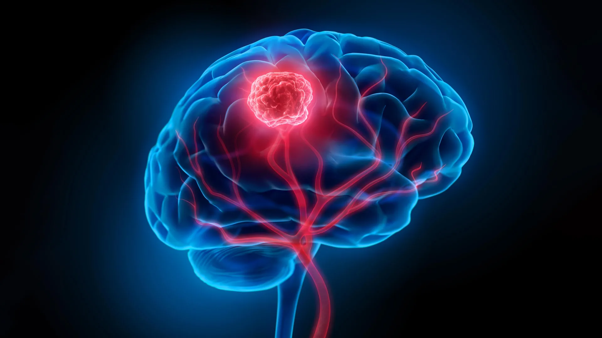
Glioblastoma, classified as a Grade IV astrocytoma, represents the most virulent and commonly diagnosed primary malignant brain tumor in adults. Its relentless progression and devastating impact on patients underscore an urgent, unmet clinical need for more effective therapeutic strategies. Despite aggressive multimodal treatment regimens that typically involve maximal surgical resection, followed by radiation therapy and adjuvant chemotherapy with temozolomide, the prognosis for individuals diagnosed with glioblastoma remains exceptionally grim. The median survival rate hovers around 15 months, with a disheartening five-year survival rate of approximately 7 percent, highlighting the profound limitations of current medical interventions.
A primary challenge in managing glioblastoma stems from its inherently infiltrative nature. Unlike many other tumors that form well-defined masses, glioblastoma cells possess a notorious capacity to disseminate diffusely into surrounding healthy brain parenchyma, often miles beyond the discernible tumor margin. This microscopic infiltration renders complete surgical extirpation virtually impossible without incurring catastrophic neurological damage. Surgeons are frequently compelled to leave behind these insidious microscopic disease foci, which inevitably serve as progenitors for tumor recurrence. Compounding this surgical dilemma is the formidable blood-brain barrier (BBB), a highly selective physiological interface that meticulously regulates the passage of substances from the bloodstream into the central nervous system. While crucial for protecting the delicate brain environment, the BBB simultaneously acts as an impenetrable shield, severely impeding the delivery of systemically administered chemotherapeutic agents and even limiting the efficacy of radiation therapy by reducing drug concentrations within the tumor microenvironment. These combined biological and anatomical hurdles are central to glioblastoma’s notorious resistance to conventional treatments and its high rates of recurrence.
In a significant stride towards surmounting these persistent obstacles, a collaborative research endeavor involving scientists from the University of Technology Sydney (UTS), Harvard University, and Henan University has unveiled a novel "double-punch" nanotherapeutic platform. Detailed in a recent publication in Science Translational Medicine, this innovative system employs a single, sophisticated material designed to address both the precision imaging requirements during surgery and the subsequent eradication of residual cancer cells. Dr. Bingyang Shi, a Chair Professor of nanomedicine from the School of Electrical, Mechanical and Biomedical Engineering at UTS, emphasized the integrated functionality of their creation, stating, "We have engineered a singular compound capable of performing two distinct, sequential tasks. It serves as an incredibly accurate guide for surgical teams during the operative phase, and subsequently transitions into a targeted therapeutic agent for post-operative clearance."
At the core of this advanced system lies a meticulously crafted, ultra-thin, two-dimensional sheet. This foundational material is engineered at the atomic level, with individual atoms precisely positioned using sophisticated manufacturing techniques adapted from the semiconductor industry. This atomic-scale precision is critical, endowing the material with the unique ability to switch seamlessly between its diagnostic and therapeutic roles. Both critical functions—enhanced tumor visualization and post-surgical phototherapy—are ingeniously activated by the application of the same near-infrared (NIR) light, a wavelength known for its superior tissue penetration capabilities and minimal invasiveness compared to visible light.
During the surgical phase, the nanoplatform transforms into a highly sensitive imaging agent, providing unparalleled clarity to neurosurgeons. The nanosheet is impregnated with a fluorescent dye specifically engineered to emit light when exposed to a specific near-infrared wavelength. This emitted fluorescence, invisible to the naked eye, allows surgeons to visualize extremely minute clusters of tumor cells, as small as 44 micrometers in diameter. To put this into perspective, conventional clinical imaging modalities, such as magnetic resonance imaging (MRI) or computed tomography (CT) scans, typically struggle to resolve structures smaller than a few millimeters. The ability to detect sub-millimeter tumor foci represents a monumental leap in surgical precision, enabling more thorough resections. Furthermore, a specialized targeting molecule is chemically conjugated to the nanosheet. This molecular appendage facilitates the selective passage of the nanoplatform across the previously impenetrable blood-brain barrier and directs its preferential accumulation within glioma cells, ensuring that the imaging agent precisely highlights cancerous tissue while minimizing signal from healthy brain regions. This unprecedented resolution empowers surgeons to delineate the tumor boundaries with far greater accuracy, potentially allowing for more aggressive yet safer removal of diseased tissue.
Following the successful excision of the macroscopically visible tumor mass, the innovative material’s therapeutic capabilities are brought to bear. The same nanosheets are carefully administered into the surgical cavity, targeting any microscopic cancer cells that inevitably remain after even the most meticulous resection. Once again, the material is activated by the very same near-infrared light. This activation triggers a sophisticated two-pronged attack on residual tumor cells. Firstly, the platinum atoms embedded within the nanosheet act as catalysts, converting the tumor’s endogenously produced hydrogen peroxide into oxygen. This process is critically important because glioblastoma, like many aggressive cancers, thrives in a profoundly hypoxic, or low-oxygen, microenvironment. This oxygen-deprived state not only promotes tumor growth and invasiveness but also confers resistance to conventional treatments like radiation and chemotherapy, which often rely on the presence of oxygen to generate cytotoxic reactive species. By effectively re-oxygenating the tumor microenvironment, the nanosheets sensitize the remaining cancer cells to subsequent therapeutic insults.
Simultaneously, the near-infrared light activation triggers two distinct phototherapeutic mechanisms: photothermal therapy (PTT) and photodynamic therapy (PDT). The light energy absorbed by the nanosheets is converted into heat, directly inducing localized hyperthermia that ablates cancer cells in their vicinity (PTT). Concurrently, the light energy also generates highly reactive oxygen species (ROS), such as singlet oxygen, within the tumor cells (PDT). These potent free radicals inflict oxidative damage to cellular components, including DNA, proteins, and lipids, ultimately leading to irreversible cell death. The synergistic action of hypoxia reversal, localized heat generation, and reactive oxygen species production creates a highly hostile environment for the surviving microscopic glioblastoma cells, providing a robust post-operative "clean-up" mechanism designed to eliminate the seeds of future recurrence.
The efficacy of this pioneering approach was rigorously evaluated in preclinical glioblastoma mouse models, yielding profoundly encouraging results. The nanoparticle treatment significantly curtailed tumor recurrence rates following surgical intervention. Notably, every single mouse in the treated cohort remained alive at the 60-day mark, a stark contrast to the control group, which received surgery alone and exhibited a median survival of only 42 days. Furthermore, comprehensive follow-up neurological and motor function assessments revealed no detectable impairments associated with the experimental treatment, suggesting a favorable safety profile in the animal subjects. These outcomes provide compelling evidence for the platform’s potential to dramatically improve post-surgical outcomes and extend survival in glioblastoma patients.
While these findings represent a beacon of hope in the challenging landscape of neuro-oncology, the research team prudently emphasizes the early-stage nature of their work. Professor Shi underscored this crucial distinction: "The results are exceptionally promising, but it is imperative to remember that this is foundational research, conducted exclusively in animal models, not yet in human subjects. This distinction holds significant weight." The transition from successful animal studies to human clinical trials is a long, arduous, and highly regulated process, fraught with numerous challenges. The performance of the imaging and therapeutic components, particularly regarding their scalability and safety, will require extensive validation within the vastly more complex environment of the human brain.
The broader field of nanomedicine has long recognized the transformative potential of engineered nanomaterials for addressing complex diseases like cancer. Nanoparticles, by virtue of their diminutive size and tunable properties, offer unprecedented opportunities for targeted drug delivery, enhanced imaging, and multimodal therapies. This glioblastoma nanoplatform exemplifies the theranostic paradigm, seamlessly integrating diagnostic and therapeutic functionalities into a single agent, thereby offering a more personalized and precise approach to disease management. Future research will focus on meticulously evaluating the long-term safety, optimal dosing, and potential for combination therapies with existing treatment modalities.
Should this groundbreaking technology continue to demonstrate its robustness and safety through subsequent rigorous testing and clinical development, the implications for glioblastoma patients could be profound. The vision is for neurosurgeons to possess an unparalleled capacity to visualize virtually every vestige of tumor during an operation and then effectively neutralize any remaining microscopic disease. This integrated strategy holds the promise of dramatically reducing the disheartening rates of tumor recurrence, which currently stand as one of the most formidable barriers to improving the survival and quality of life for individuals grappling with glioblastoma. This advancement signifies a meaningful stride towards a future where glioblastoma might be managed with far greater precision and efficacy.
About the Author