Congenital heart disease (CHD) represents the most prevalent form of birth defect globally, impacting approximately one in every hundred newborns annually. These structural abnormalities of the heart, present from birth, can range from mild to life-threatening, often requiring complex medical interventions and lifelong management. While numerous genetic and environmental factors contribute to CHD, alterations in specific genes are frequently implicated. Among these, the TBX5 gene has long been recognized for its crucial role in orchestrating the development of a healthy heart. Typically, individuals inherit two functional copies of each gene, one from each parent. However, in certain instances of CHD, a child possesses only a single functional copy of TBX5, with the other either missing or non-functional.
For an extended period, the precise molecular mechanisms by which the loss of just one functional TBX5 gene copy — a phenomenon known as haploinsufficiency — could precipitate such profound developmental anomalies in the heart remained a significant enigma for geneticists and developmental biologists. Despite the presence of one perfectly healthy copy, its sufficiency was often questioned, leading to an incomplete understanding of disease pathology. This persistent question has driven extensive research into how gene dosage, specifically the reduction to half the normal level of a critical protein, can exert such a dramatic impact on cellular processes and organ formation.
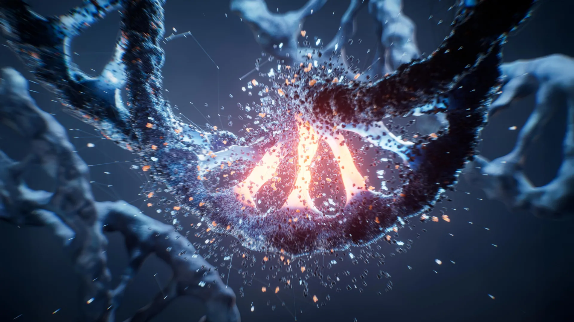
A groundbreaking study published in the esteemed journal Science by researchers at Gladstone Institutes has now provided a compelling answer, fundamentally shifting our understanding of TBX5’s function and, by extension, the etiology of certain congenital disorders. This new research reveals that TBX5 performs a vital function beyond its well-established role in regulating gene expression. It acts as a critical organizer, meticulously arranging the complex, three-dimensional physical structure of DNA within heart cells – an architectural role indispensable for proper cardiac function. The scientists discovered that even the reduction to a single functional copy of TBX5 is enough to severely disrupt this intricate genomic organization, consequently altering the activation patterns of a multitude of other genes essential for heart development.
This revelation offers an entirely novel perspective on the long-standing genetic puzzle of haploinsufficiency, explaining why the insufficient quantity of certain proteins, even when other copies are intact, can lead to severe developmental issues. Dr. Benoit Bruneau, Director of the Gladstone Institute of Cardiovascular Disease and a senior author of the study, emphasized the broader implications: "TBX5 serves as an illustrative case for a wider category of genes that, when only one copy is lost, are associated with various birth defects. What makes our findings particularly compelling is the suggestion that a common underlying mechanism — the incorrect folding of the cell’s three-dimensional genetic blueprint — might explain many distinct developmental anomalies." Dr. Katie Pollard, Director of the Gladstone Institute of Data Science and Biotechnology and the other senior author, highlighted the computational prowess required: "We leveraged and developed sophisticated computational models to meticulously analyze data from thousands of individual cells. This enabled us to finally visualize how the diminished presence of this crucial protein triggers a systemic breakdown in the heart’s DNA structure across every hierarchical level."
To truly appreciate the significance of these findings, it’s essential to understand the remarkable feat of DNA packaging within a cell. The human genome, an instruction manual stretching over two meters in length, must be precisely compacted into a microscopic nucleus, akin to condensing an epic novel into a thimble. This isn’t a random process; each cell type, whether a heart cell, a brain cell, or a skin cell, organizes its genetic material into a unique, highly specific three-dimensional configuration. This cell-specific folding dictates which genes are accessible and active, thereby determining the cell’s identity and function.
This intricate 3D genomic architecture is structured hierarchically. It includes large, distinct compartments that can be thought of as separate volumes of a manual, further subdivided into topologically associating domains (TADs), analogous to chapters or paragraphs. At an even finer scale are chromatin loops, where distant segments of DNA are brought into close physical proximity. These loops are particularly vital, enabling regulatory elements called enhancers – genetic switches located far from the genes they control – to physically interact with their target genes. Such physical contacts are crucial for activating the specific genetic instructions required for a cell to develop and function correctly.
Prior research had already established TBX5 as a key transcriptional regulator, meaning it directly influences the expression of many genes essential for the development and proper functioning of heart cells. Earlier investigations from Dr. Bruneau’s laboratory had indicated that a reduction in TBX5 levels affected the expression of hundreds of other heart-specific genes. However, the precise mechanism linking TBX5 reduction to altered gene expression and, ultimately, heart defects remained elusive. The research team therefore embarked on an ambitious endeavor to ascertain whether the physical organization of DNA directly influences heart cell behavior, and whether TBX5 played a controlling role in this architectural process.
To conduct their investigation, the scientists employed a sophisticated combination of advanced methodologies, enabling them to scrutinize individual cellular responses to varying concentrations of TBX5. They began by guiding human induced pluripotent stem cells – cells capable of differentiating into any cell type – to mature into beating heart muscle cells. These cultures were established under three conditions: healthy cells with two functional TBX5 copies, cells engineered to lack one TBX5 copy (modeling haploinsufficiency), and cells completely devoid of TBX5. Subsequently, the team utilized high-resolution three-dimensional genomic mapping techniques to meticulously examine the formation and structure of DNA loops within these cells at an unprecedented level of detail.
Given the immense scale of the experiment, which generated millions of data points from thousands of individual cells, the researchers relied heavily on custom-developed computational models to process and interpret the vast datasets. "Through the tailored computational approaches we engineered, we were able to observe for the first time how the diminished presence of TBX5 initiates a widespread disintegration of the heart’s 3D DNA organization," stated Dr. Shuzhen Kuang, a first author of the study and a former bioinformatics fellow in Dr. Pollard’s lab. "Remarkably, this structural breakdown was evident across all organizational scales of the genome – from the broad compartments to the more localized domains and the critical chromatin loops."
The study definitively demonstrated that TBX5 functions as a central architect during the maturation of heart cells. As healthy stem cells progressed into specialized cardiac muscle cells, the researchers observed profound and dynamic reorganizations of the genome. Extensive segments of DNA transitioned between active and inactive states, reflecting the precise gene expression programs required for cellular differentiation. TBX5 emerged as a pivotal orchestrator of these structural transformations.
The team uncovered that TBX5 acts much like a cellular navigation system, guiding a crucial molecular motor protein known as cohesin. TBX5 directs cohesin to specific locations on the DNA, where it then facilitates the formation of chromatin loops. These loops are essential for bringing distant genes into direct contact with their corresponding enhancers, thereby enabling their activation. When TBX5 levels are insufficient, this navigational system falters. Cohesin is misdirected, or its activity is impaired, leading to improper chromatin loop formation. Consequently, the DNA becomes incorrectly folded, and vital genes responsible for cardiac development fail to be activated at the precise moments they are needed, directly contributing to the formation of heart defects.
"What was truly striking was the critical importance of TBX5 dosage," commented Dr. Zoe Grant, a first author of the study and a postdoctoral researcher in Dr. Bruneau’s lab. "The greater the reduction in TBX5, the more severe and pervasive the disruption became across every level of genome organization we investigated." The findings unequivocally showed that merely halving the normal quantity of TBX5 is sufficient to trigger widespread DNA misfolding and directly contribute to the genesis of congenital heart defects.
The research also brought to light another intriguing aspect: individual heart cells do not exhibit identical responses to the loss of TBX5. Notable distinctions were observed between the two primary types of heart cells, atrial and ventricular cells, which have different developmental trajectories and functions. Furthermore, even among cells of the same type, a degree of variation was detected. "This cellular heterogeneity could offer an explanation for why individuals carrying the identical genetic mutation might present with a spectrum of different heart defects, ranging in type and severity," Dr. Grant suggested.
While these discoveries provide profound new clarity into the mechanisms underpinning congenital heart disease, the researchers propose that this fundamental mechanism may extend to other developmental disorders. "We believe we have uncovered a novel mechanism of disease pathogenesis," Dr. Bruneau asserted. "Our work demonstrates that even a modest reduction in a single protein can cause the genomic blueprint to fold incorrectly, leading directly to disease. Therefore, many birth defects currently attributed solely to genetic mutations might, in fact, be rooted in the three-dimensional misfolding of DNA." This implies a paradigm shift, suggesting that some genetic mutations cause disease not merely by altering individual genetic instructions but by fundamentally disturbing the physical arrangement and accessibility of the entire genome.
Looking ahead, the research team intends to pinpoint the precise developmental stage at which TBX5 initiates its critical genome organizing function during early cardiac formation. They also aim to investigate whether other proteins implicated in various birth defects exert similar architectural control over DNA structure, potentially uncovering a broader class of disorders linked to this novel mechanism. The comprehensive findings of this study, "Dose-dependent sensitivity of human three-dimensional chromatin to a heart disease-linked transcription factor," published on July 23, 2026, were the result of a collaborative effort involving researchers from Gladstone, UC San Diego, and UC San Francisco, and received support from various prestigious institutions including the National Institutes of Health. This work marks a significant stride in understanding the complex interplay between genes, their architectural roles, and human development.
About the Author