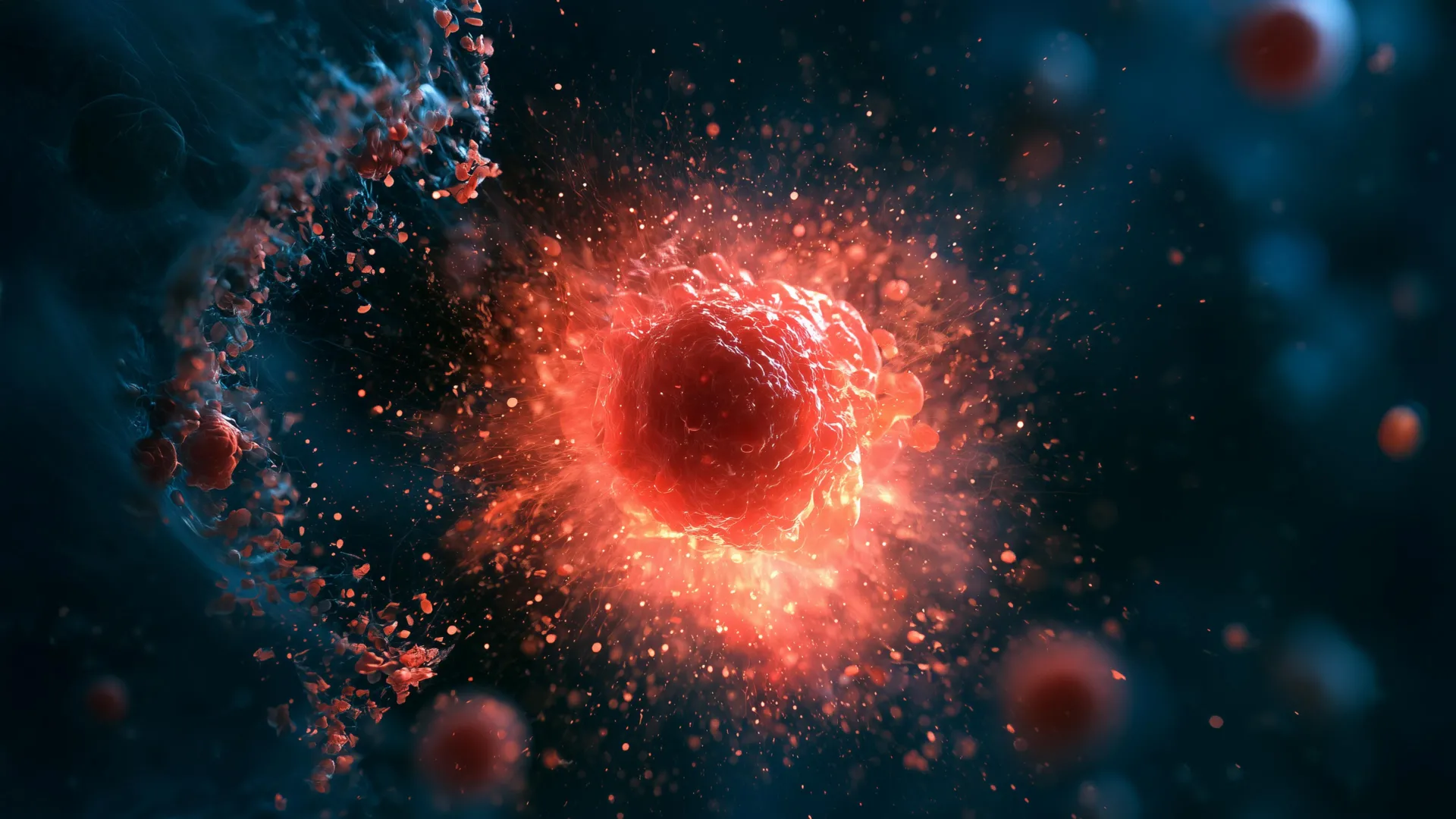
Stanford University researchers have unveiled a novel class of immune cells, dubbed "ruptoblasts," possessing an extraordinary and hitherto unobserved mechanism for eliminating threats: a self-destructive detonation that eradicates both the cell itself and its immediate surroundings. This dramatic cellular event, characterized by an explosive release of internal contents, results in the cell’s complete disintegration within mere minutes, leaving behind minimal residual evidence. The groundbreaking discovery, detailed in a recent publication in the esteemed journal Cell, emerged from an in-depth investigation into the regenerative capabilities of planarian flatworms, an ancient lineage of aquatic invertebrates renowned for their remarkable capacity to regrow damaged or missing body parts, and in certain instances, to generate entire new organisms from fragments. The evolutionary persistence and adaptation of the immune systems in these creatures over hundreds of millions of years offer a tantalizing window into potential new therapeutic avenues for human medicine.
The research team, led by associate professor of bioengineering Bo Wang, initially embarked on a quest to understand how flatworms differentiate between their own tissues and foreign invaders. Postdoctoral researcher Chew Chai, a key figure in the study, observed these peculiar cellular responses while meticulously examining the immune reactions of fused flatworms. To probe the worms’ ability to recognize self versus non-self, Chai performed experiments involving longitudinal incisions and subsequent fusion with segments from unrelated individuals. While flatworms possess an innate proficiency in tissue repair and regeneration, the fused specimens exhibited a distinct rejection response towards tissue sourced from different worms, a phenomenon that bears a conceptual resemblance to the immune rejection of transplanted organs in humans, albeit with a vastly different cellular execution.
The observed reaction in the flatworms was unlike any previously documented immune process. Chai described it as an intense inflammatory cascade, akin to an urgent alarm triggering a widespread cellular detonation. This profound inflammatory state was found to be directly influenced by the hormone activin. Prior research had established activin’s crucial role in flatworm biology, with elevated levels hindering regeneration and insufficient levels impacting reproductive capabilities. As the fused worms initiated their rejection of foreign tissue, Chai noted a concomitant surge in activin, which precipitated a sustained inflammatory response. Although the affected worms did not succumb immediately, their survival was curtailed to a matter of days. Intriguingly, the injection of activin into healthy, unfused flatworms elicited a comparable inflammatory reaction, underscoring activin’s role as a potent trigger for this defensive mechanism.
To dissect the precise cellular events underlying this explosive response, Chai employed sophisticated live-cell microscopy and flow cytometry, a technique utilizing laser-based analysis to identify and sort cells. By labeling cells with fluorescent markers, she was able to isolate and observe those cells that responded to activin. The pivotal observation was the sudden and violent rupture of a specific subset of cells. These cells, upon activation, unleashed their internal contents, causing the demise of adjacent cells, and then rapidly vanished. This explosive cell death process was christened "ruptosis," and the cells responsible were termed "ruptoblasts."
The defining characteristic of ruptosis is its extraordinary speed and completeness, setting it apart from other known modes of programmed cell death. While some mammalian cells and bacteria may exhibit forms of explosive cell death, these processes typically unfold over extended periods, often hours, involving a gradual leakage of cellular components. In stark contrast, ruptosis occurs within seconds to minutes, transforming the ruptoblast into an immediate and potent effector of localized destruction. This rapid expulsion of cellular contents functions as a highly targeted weapon, delivering a decisive blow rather than a slow-acting poison.
To ascertain the efficacy and scope of this defensive mechanism, the researchers subjected various targets to ruptoblast activity. Experiments demonstrated that ruptoblasts could effectively destroy E. coli bacteria, human kidney cells, and mouse blood cells. Crucially, the destructive impact was confined to cells in immediate proximity to the ruptoblast. The process did not instigate a chain reaction of cell death nor did it leave behind persistent toxic residues. According to Professor Wang, this capacity for a powerful yet precisely contained assault holds significant promise for the development of future therapeutic strategies aimed at combating bacterial infections or targeting cancerous growths.
Ruptoblasts diverge significantly from more commonly studied immune cells such as T cells and neutrophils, which are hematopoietic in origin, meaning they are derived from bone marrow. Ruptoblasts, conversely, are glandular cells, suggesting a different developmental pathway and functional specialization. The researchers hypothesize that ruptoblasts augment their standard secretory mechanisms to enable the sudden and forceful expulsion of toxic substances upon encountering activin. A rapid influx of calcium ions from the endoplasmic reticulum within the cell is believed to be a critical driver of the ruptosis cascade.
The distribution of ruptoblasts appears to be ancient, with similar cells identified only in basal bilaterians, including flatworms. This restricted prevalence suggests that ruptoblasts represent an early evolutionary innovation in immune defense. Chew Chai posits that vertebrates may have evolved away from this strategy due to the inherent difficulty in repairing the collateral damage inflicted by ruptosis. In contrast, flatworms, with their abundant stem cell populations and exceptional regenerative abilities, are well-equipped to replace tissues damaged by such potent defensive actions.
Professor Wang emphasized the broader implications of this discovery, highlighting the vast diversity of immune mechanisms that exist across the animal kingdom, many of which remain largely unexplored. The study of organisms less commonly employed as traditional research models, like flatworms, has the potential to reveal novel immune strategies absent in humans and other vertebrates, thereby expanding our understanding of biological defense systems. Examining a wider array of species, Wang suggested, could spark innovative approaches to some of the most intractable challenges in modern medicine. The findings underscore the immense value of investigating the intricate biology of seemingly simple organisms to unlock profound biological insights.
The research was supported by a multitude of prestigious grants and fellowships, including funding from the National Science Foundation Graduate Research Fellowship, a Stanford Graduate Fellowship, a Stanford DARE fellowship, a Human Frontier Science Program grant, a National Institutes of Health grant, and the European Research Council. The collaborative effort involved numerous researchers from Stanford University, including postdoctoral scholar Souradeep Sarkar, former Undergraduate Visiting Research Program scholar Lihan Zhong, Dania Nanes Sarfati, Christine Jacobs-Wagner, and Hawa Racine Thiam. Additional contributions came from co-senior author Benyamin Rosental and his team at Ben Gurion University of the Negev. Several researchers involved hold affiliations with prominent interdisciplinary centers at Stanford, including Stanford Bio-X, the Wu Tsai Neurosciences Institute, and Sarafan ChEM-H, as well as the Maternal & Child Health Research Institute (MCHRI).
About the Author