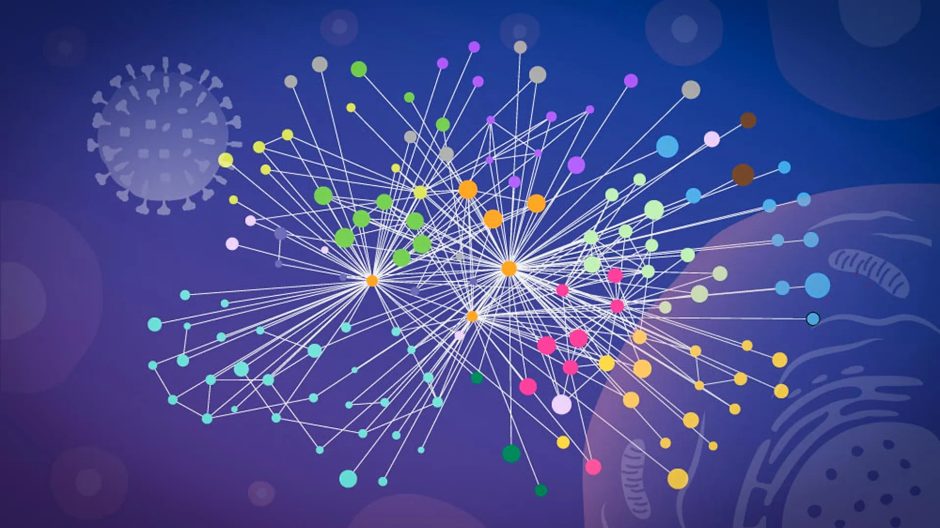
A groundbreaking advancement in understanding the intricate strategies employed by influenza A to commandeer human cellular machinery has been unveiled, thanks to a collaborative effort by researchers at the European Molecular Biology Laboratory (EMBL) Hamburg and the Leibniz Research Institute for Molecular Pharmacology (FMP). This novel research has generated an unprecedentedly detailed molecular map, not by dissecting cells, but by observing viral and host protein interactions in their natural, intact cellular environment. This innovative approach circumvents the limitations of traditional methods that often disrupt the delicate molecular architecture of an infected cell, thereby providing a more accurate and nuanced portrayal of viral pathogenesis.
The global burden of seasonal influenza remains significant, with millions of severe illnesses and hundreds of thousands of fatalities annually, underscoring the persistent threat posed by this ubiquitous pathogen. Furthermore, influenza A has been the architect of devastating pandemics throughout history, most notably the 1918 Spanish Flu, which dramatically reshaped global health landscapes. The fundamental modus operandi of the influenza virus involves its entry into a host cell, followed by the release of its genetic material, RNA, which carries the blueprints for synthesizing a limited repertoire of viral proteins. These proteins then infiltrate the host cell, subtly re-engineering its internal molecular systems and transforming the cell into a highly efficient factory dedicated to the production of new viral progeny.
Gaining a comprehensive understanding of this cellular takeover is paramount for the development of more robust influenza vaccines and more effective antiviral therapies. This necessitates precise knowledge of which viral proteins engage with human proteins, the specific cellular locations where these critical interactions occur, and the precise ways in which the virus exploits these connections to facilitate its own replication cycle. The current study marks a significant milestone as the first large-scale mapping of direct physical contacts between influenza viral proteins and human host proteins within intact, infected cells. The level of structural resolution achieved is so refined that it allows for the computational modeling of how these interacting protein partners are likely to physically fit together.
Jan Kosinski, a Group Leader at EMBL Hamburg and the Centre for Structural Systems Biology (CSSB), emphasized the transformative nature of their work, stating that it introduces a novel paradigm for examining flu-host interactions within their native context, enriched with invaluable structural insights. He further elaborated that the current findings represent a static snapshot of a particular phase during the infection process, but crucially, they pave the way for a dynamic exploration of flu-host interactions across the entirety of the viral life cycle within the cell.
The inherent difficulty in tracking protein-protein interactions during an active infection has historically presented a formidable experimental hurdle. Many prior investigations relied on biochemical techniques that necessitated the complete disruption of cellular integrity before protein contacts could be quantified. This disruptive process inherently risks distorting the natural state of affairs within a living cell. Once cellular compartments are dismantled, proteins that were once spatially segregated might come into proximity in the artificial laboratory setting. Concurrently, transient, weak, or location-specific interactions, which could be vital for viral function, might vanish entirely. Consequently, researchers have often faced challenges in discerning which molecular connections are genuine occurrences during infection and which are merely artifacts of the experimental procedure.
It was in this challenging research landscape that the team discovered that their collaborators, Boris Bogdanow and Fan Liu, at the FMP in Berlin, had ingeniously adapted a well-established technique known as cross-linking mass spectrometry (XL-MS). This specialized adaptation was meticulously tailored for the specific demands of studying virus-infected cells, providing the critical breakthrough the researchers had been seeking. This refined methodology enabled the capture of protein interactions that are fleeting in duration or confined to particular subcellular regions within an infected cell.
Boris Bogdanow, now a Junior Research Group Leader at the Institute of Virology at Charité – Universitätsmedizin Berlin, explained that XL-MS permits the direct capture of protein-protein interactions within intact, infected cells, while simultaneously furnishing structural information regarding the nature of these interactions. He further elaborated that this dual capability offers profound insights into the interface between the virus and the human cell, with the potential, through structural modeling, to pinpoint actionable targets for future pharmaceutical interventions.
The researchers strategically combined their experimental XL-MS data with sophisticated computational structural modeling techniques. This synergistic approach enabled them to not only identify specific viral and human proteins that engage in direct contact but also to estimate their relative spatial orientations at the moment of interaction. To construct these detailed structural models, the team employed a modified iteration of AlphaFold, the renowned protein structure prediction algorithm that garnered a Nobel Prize.
The principal advantage of this modified AlphaFold approach, according to Kosinski, lies in its capacity to directly incorporate the experimentally derived cross-linking data into the structural modeling process. This integration effectively guides the model by informing it about the proximity of specific regions of viral and host proteins within the infected cell. This feature proved particularly valuable for analyzing virus-host complexes, which are notoriously challenging to predict with high accuracy using conventional methods.
The findings, published in the esteemed journal Nature Microbiology, have illuminated at least two distinct and significant strategies that influenza A virus appears to employ to assert control over a host cell. The first key discovery centers on hemagglutinin, a critical protein situated on the exterior surface of the influenza virus, which serves as the primary mechanism for the virus to attach to and subsequently enter host cells. The research team meticulously tracked the journey of hemagglutinin as it navigated the cell’s complex internal transport and processing network. This intricate network comprises various membrane-bound compartments responsible for folding, modifying, and preparing proteins before directing them to their designated functional locations. The detailed analysis revealed that a surprising number of human proteins actively participated in the correct folding and modification of hemagglutinin during the course of infection. Intriguingly, some of these host proteins had previously been associated with poorly understood cellular functions, highlighting the virus’s ability to co-opt even less-characterized cellular machinery.
The second major revelation from the study pertains to paraspeckles, which are small, droplet-like structures located within the nucleus of the cell. The research team observed a consistent and striking phenomenon: influenza A infection induced the dissolution of these paraspeckles. Upon the disintegration of paraspeckles, they released their cargo of RNA-binding proteins, which had been sequestered within these structures. The virus then appears to strategically repurpose these liberated proteins to facilitate its own replication processes.
Iuliia Kotova, a former predoctoral fellow in the Kosinski Group at EMBL Hamburg and the first author of the publication, expressed her astonishment at the observation of paraspeckles, stating that witnessing these minute nuclear organelles consistently dissolve across all tested cell lines and influenza strains suggested that this disruption was not merely an incidental consequence of infection but rather a deliberate viral strategy. The implications of this paraspeckle disruption extend beyond the mere release of RNA-binding proteins. Kosinski further speculated that there might be a secondary benefit for the virus, as some evidence indicates that paraspeckles play a role in cellular stress responses and the regulation of antiviral genes. Therefore, their disruption could simultaneously compromise the cell’s intrinsic defense mechanisms.
The success of this ambitious project was contingent upon the synergistic pooling of specialized technologies and scientific expertise across three distinct institutions. The critical cross-linking mass spectrometry analyses were conducted at Charité in Berlin. Comprehensive glycoproteomics analyses, which examine the structure and function of proteins with attached carbohydrates, were performed at the EMBL Proteomics Core Facility. The complex computational structural modeling, leveraging the modified AlphaFold algorithm, was executed on the powerful EMBL Compute Cluster. Concurrently, advanced microscopy imaging, essential for visualizing cellular structures and events, took place at the Advanced Light and Fluorescence Microscopy (ALFM) Facility at CSSB.
This pioneering study unequivocally demonstrates the profound value of investigating molecular interactions within the context of intact infected cells. Such an approach offers unparalleled insights into both the specific locations and the precise mechanisms by which viruses subvert human cellular machinery. The researchers posit that this "mapping in context" methodology holds significant potential for deciphering the operational strategies of a wide array of other viruses.
Kosinski articulated that while the precise host factors and mechanistic pathways may vary considerably between different viruses, the overarching approach – which integrates in-cell cross-linking, structural modeling, and targeted cell-biology investigations to map native virus-host interactions at specific stages of infection – remains broadly applicable. Although the current investigation utilized a laboratory-adapted strain of influenza, the researchers are optimistic that this same methodological framework can be extended to scrutinize viruses with a higher potential for pandemic emergence. Bogdanow concurred, noting that while this study focused on a lab-adapted strain, it lays a robust foundation for applying this methodology to viruses of significant pandemic relevance, such as H5N1, thereby facilitating the uncovering of the intricate interaction networks that underpin their propagation within human cells.
About the Author