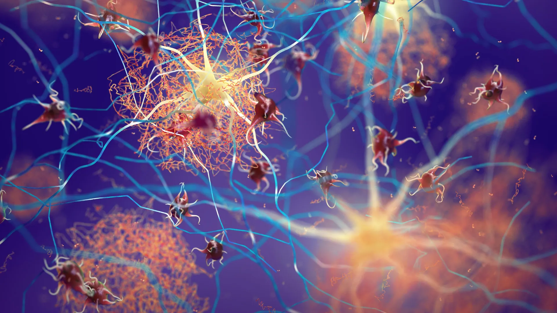
Groundbreaking research originating from Baylor College of Medicine has illuminated a previously underappreciated cellular component, tubulin, as a potential linchpin in the fight against neurodegenerative conditions such as Alzheimer’s and Parkinson’s disease. These devastating illnesses are intrinsically linked to the pathological aggregation of specific proteins within the brain, namely Tau and alpha-synuclein. The conventional understanding has largely focused on these proteins’ propensity to misfold and coalesce into toxic clumps, which disrupt neuronal function and ultimately lead to cognitive and motor impairments. However, this new investigation, detailed in the esteemed journal Nature Communications, proposes a paradigm shift, identifying tubulin not merely as a structural element but as an active modulator that can steer these proteins away from destructive pathways.
The cellular architecture of neurons relies heavily on a sophisticated internal scaffolding system known as microtubules. These dynamic structures, akin to a complex network of internal highways, are crucial for transporting vital cellular components, maintaining cell shape, and facilitating intracellular communication. Tubulin, a globular protein, serves as the fundamental building block of these microtubules. The research team has posited and demonstrated that the presence and abundance of tubulin play a pivotal role in dictating the fate of Tau and alpha-synuclein. Rather than merely preventing the formation of aberrant protein structures, tubulin appears to actively engage these proteins, channeling them towards their essential physiological functions within healthy neuronal environments. This redirection mechanism could offer a powerful therapeutic avenue, distinct from approaches that aim to eliminate the protein aggregates entirely, which might inadvertently compromise normal cellular processes.
The accumulation of misfolded Tau and alpha-synuclein proteins is a hallmark of numerous neurodegenerative disorders. In Alzheimer’s disease, Tau proteins undergo modifications that cause them to detach from microtubules and aggregate into neurofibrillary tangles, disrupting neuronal transport and leading to cell death. Similarly, Parkinson’s disease is characterized by the formation of Lewy bodies, primarily composed of misfolded alpha-synuclein, which damage dopamine-producing neurons in the substantia nigra, resulting in the characteristic motor symptoms. "These proteins, Tau and alpha-synuclein, are profoundly implicated in the pathogenesis of neurodegenerative diseases like Alzheimer’s and Parkinson’s," explained Dr. Lathan Lucas, the study’s lead author and a postdoctoral associate in the lab of Dr. Allan Ferreon. "Their tendency to misfold and aggregate into detrimental structures is a primary driver of neuronal damage, manifesting as memory deficits, motor control issues, and a spectrum of other debilitating symptoms."
However, the narrative surrounding Tau and alpha-synuclein is far more nuanced than their disease-causing potential. In healthy, functioning neurons, these proteins are not inherently detrimental; rather, they fulfill critical roles that are indispensable for neuronal viability and communication. They are integral to maintaining cellular integrity and play a significant part in the intricate processes of microtubule assembly and stabilization. "It’s crucial to recognize that Tau and alpha-synuclein also perform vital functions within healthy neurons," Dr. Lucas elaborated. "They contribute to cell structure and are instrumental in supporting neuronal communication, largely through their interactions with tubulin and their involvement in the dynamic construction and maintenance of microtubules."
A key area of focus in the study was the concept of cellular condensates. These are not membrane-bound organelles but rather dynamic, liquid-like droplets that form within the cell cytoplasm. They are believed to concentrate specific proteins and RNA molecules, thereby regulating various cellular processes. Both Tau and alpha-synuclein have been observed to form these condensates, and their aberrant behavior within these structures is thought to be a critical step in the cascade leading to protein aggregation and disease. Consequently, disrupting the formation of these condensates has been a proposed therapeutic strategy. Yet, this approach carries inherent risks. Given that condensates also play crucial roles in normal brain function, indiscriminately eliminating them could have unintended and detrimental consequences for healthy neuronal activity.
This complex interplay between beneficial and harmful functions within condensates led the researchers to a novel hypothesis. Instead of attempting to dismantle these cellular micro-environments, what if the focus could be shifted towards influencing the behavior of the proteins residing within them? "This contemplation led us to a critical question: rather than preventing the formation of these condensates, could we engineer conditions that would guide Tau and alpha-synuclein towards their beneficial roles, thereby discouraging their deviation towards disease-associated pathways?" posed Dr. Allan Ferreon, an associate professor and co-corresponding author of the study.
To illustrate this concept, Dr. Lucas employed a relatable analogy. "Imagine Tau and alpha-synuclein as potentially disruptive students in a classroom," he suggested. "One approach is to isolate them and give them little to do, which might lead to them acting out. Alternatively, one can engage them constructively in academic pursuits, sports, or creative activities, channeling their energy productively and preventing them from causing trouble. Our findings suggest that tubulin acts in a similar fashion, effectively directing these proteins towards a constructive, healthy path."
The research team meticulously investigated this hypothesis through a sophisticated combination of biochemical and biophysical techniques, augmented by high-resolution microscopy and experiments conducted in living neurons. Their primary objective was to ascertain whether tubulin could indeed influence the behavior of Tau and alpha-synuclein, specifically to prevent the formation of toxic aggregates within cellular condensates.
The experimental findings strongly supported their hypothesis. The study revealed a significant correlation between tubulin levels and the propensity for Tau and alpha-synuclein to form toxic aggregates. "We observed that when tubulin levels are diminished, a condition that has been noted in some neurodegenerative diseases, the microtubules become less abundant, creating an environment where Tau and alpha-synuclein are more likely to misfold and aggregate into toxic structures," Dr. Lucas explained.
Conversely, the presence of sufficient tubulin acted as a protective factor. "When tubulin is readily available, Tau and alpha-synuclein are nudged away from forming harmful aggregates. Instead, they are encouraged to participate in the assembly of healthy microtubules," he continued. "Tubulin effectively reorients the activity of these proteins by providing them with a productive outlet for their cellular functions." This mechanism suggests that tubulin is not merely a passive bystander but an active participant in maintaining neuronal health.
The implications of these findings are substantial, suggesting a far more active and protective role for tubulin in brain health than previously recognized. "Our discoveries fundamentally redefine the role of tubulin in neurodegeneration, transitioning it from a passive entity susceptible to disease processes to an active defender against the formation of toxic protein aggregates," stated Dr. Ferreon. "The potential therapeutic strategy emerging from this work is to augment the tubulin pool, rather than attempting to block the formation of cellular condensates. This approach could effectively curtail toxic aggregation while simultaneously preserving the essential physiological roles of Tau and alpha-synuclein, offering a highly selective and promising therapeutic avenue."
The collaborative effort behind this research included co-first author Phoebe S. Tsoi, My Diem Quan, Kyoung-Jae Choi, and co-corresponding author Josephine C. Ferreon, all affiliated with Baylor College of Medicine. Funding for this significant investigation was generously provided by grants from the National Institute of Neurological Disorders and Stroke (NINDS) of the National Institutes of Health (NIH) under award number R01 NS105874, the Welch Foundation under grant Q-2097-20220331, and the National Institute of General Medical Sciences (NIGMS) of the NIH under award number R01 GM122763.
About the Author