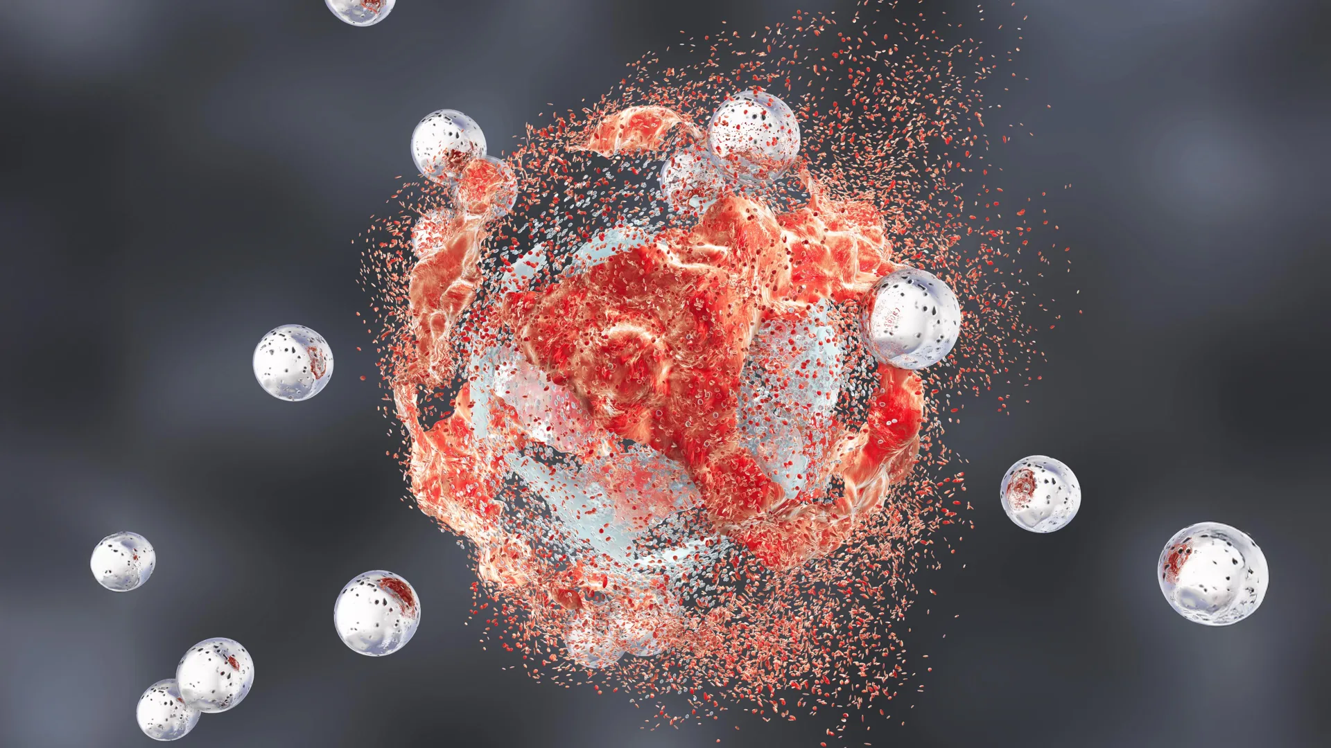
Prostate cancer remains a significant health challenge globally, with aggressive forms posing particular difficulties in treatment due to their metastatic potential and resistance to conventional therapies. A groundbreaking preclinical investigation, spearheaded by scientists at Weill Cornell Medicine and the Cornell Duffield College of Engineering, has unveiled a novel therapeutic approach utilizing ultrasmall silica nanoparticles that not only directly eliminate prostate tumors but also recalibrate the body’s immune system to actively combat cancerous cells. This innovative strategy, tested in mouse models of aggressive prostate carcinoma, yielded multiple instances of complete tumor regression, offering compelling evidence for its potential translation into human clinical applications.
The sophisticated nanoparticles, officially designated as ultrasmall fluorescent core-shell silica nanoparticles and known colloquially as Cornell Prime dots, or C’ dots, represent a fascinating intersection of materials science and biomedical engineering. Initially conceived and engineered to enhance the precision and clarity of medical imaging techniques, these particles have already progressed to advanced stages of clinical evaluation for applications such as image-guided surgical procedures and other diagnostic utilities. However, ongoing research unexpectedly uncovered an intrinsic therapeutic capacity within these particles: a remarkable ability to selectively damage and destroy malignant cells while largely preserving the integrity and function of healthy tissue. This serendipitous discovery has opened new avenues for their application beyond mere visualization.
The detailed findings of this study, recently published in the esteemed journal Cancer Research, a publication of the American Association for Cancer Research, illuminate the multifaceted mechanism through which these silica nanoparticles exert their anti-cancer effects. The research team observed that the particles render tumor cells exceptionally susceptible to a specific type of programmed cell death known as ferroptosis. Concurrently, the nanoparticles fundamentally alter the immunological landscape surrounding the tumor, transforming what is often an immune-resistant, or "cold," microenvironment into an immune-responsive, "hot" state. This profound shift in the tumor microenvironment (TME) is critically important, as it holds the potential to dramatically augment the efficacy of existing immunotherapeutic agents, which often struggle to achieve sustained responses in immune-cold tumors. Dr. Michelle Bradbury, the senior author of the study and the Endowed Professor of Imaging Research in Radiology and director of the Molecular Imaging Innovations Institute at Weill Cornell Medicine, emphasized the transformative potential: "These results are highly encouraging; a therapeutic modality that simultaneously triggers tumor-cell demise and reconfigures the immune microenvironment, as this does, would establish an entirely new clinical paradigm for cancer management."
The development of these pioneering nanoparticles is the culmination of an extensive and enduring collaborative effort between Dr. Bradbury’s laboratory and the research group led by Dr. Ulrich Wiesner, a co-corresponding author and the Spencer T. Olin Professor in the Department of Materials Science and Engineering at Cornell University, also affiliated with the College of Architecture, Art, and Planning. This interdisciplinary partnership, bolstered by financial support from entities such as the Parker Institute for Cancer Immunotherapy at Weill Cornell Medicine, underscores the intricate nature of advanced cancer research.
One of the most remarkable discoveries pertaining to the nanoparticles’ mechanism of action centers on their ability to induce ferroptosis. Unlike other well-known forms of programmed cell death, such as apoptosis or necrosis, ferroptosis is uniquely characterized by its dependence on iron and an overwhelming accumulation of reactive oxygen species, particularly leading to lipid peroxidation. This process severely damages the fatty molecules that constitute cellular membranes, ultimately causing the cell to rupture and disintegrate. While the precise molecular pathways by which the silica nanoparticles initiate this process are still under comprehensive investigation, compelling evidence suggests a crucial role for iron. The nanoparticles, originally designed for their imaging capabilities, appear to actively sequester positively charged iron ions from the bloodstream and transport them directly into tumor cells. Once inside the malignant cells, these concentrated iron ions are hypothesized to fuel the intense oxidative stress that is the hallmark of ferroptosis, thereby initiating the destructive cascade. This targeted delivery of a ferroptosis-inducing agent represents a highly strategic approach to cancer cell elimination.
Beyond their direct cytotoxic effects, the silica nanoparticles demonstrate an equally profound impact on the immune system surrounding the cancerous growth. Many aggressive tumors employ sophisticated mechanisms to evade immune surveillance, creating an immunosuppressive tumor microenvironment that deactivates or recruits immune cells to protect the tumor rather than attack it. This results in an immunologically "cold" tumor, notoriously resistant to immunotherapies. The researchers observed that in the presence of the nanoparticles, immune cells within the tumor’s vicinity—including T lymphocytes, macrophages, and other critical components of the immune system—underwent a significant phenotypic transformation. They shifted from an inactive or immune-suppressing state to one of robust activity and aggressive cancer-fighting capability. This re-education of the immune microenvironment makes the tumors substantially more receptive to established immunotherapy drugs, such as immune checkpoint inhibitors. Concurrently, the nanoparticles also interfered with critical metabolic processes across various cell types within the TME, further impeding tumor growth and proliferation. To ensure the precise delivery of this multi-pronged treatment specifically to prostate cancer cells, the research team ingeniously functionalized the nanoparticles with a targeting molecule designed to recognize prostate-specific membrane antigen (PSMA), a protein abundantly expressed on the surface of prostate tumor cells. While some transient accumulation of the particles was noted in other organs, such as the spleen, the comprehensive safety evaluations conducted found no indications of systemic toxicity or adverse effects on healthy tissues outside the tumors, highlighting the precision of the targeted delivery system. Dr. Wiesner expressed his astonishment at the breadth of effects: "It seems almost unbelievable—how can it be that instead of a single pathway, we observe all these diverse effects occurring simultaneously, exclusively within tumors, and without impacting healthy tissues? It makes me contemplate whether the ancient and ubiquitous presence of ultrasmall silica in our environment and in common foods like leafy greens or cereal grains has fostered a fundamental biological connection that we are only now beginning to comprehend."
The most compelling evidence of the nanoparticles’ therapeutic power emerged from survival studies conducted on mice afflicted with aggressive prostate cancer. When administered as monotherapies, both the C’ dots alone and conventional immunotherapy agents individually yielded modest improvements in survival rates compared to untreated control groups. However, the true synergistic potential became evident when the nanoparticles were combined with an immune checkpoint blockade therapy. This combination regimen resulted in complete or nearly complete tumor remissions and indefinite survival in a remarkable four out of ten treated mice, a statistically and clinically significant outcome in such aggressive cancer models. Further augmenting the therapeutic cocktail by incorporating a third treatment, a CSF-1R blockade, which specifically targets tumor-associated macrophages that often promote tumor growth and immune suppression, further enhanced these impressive results, increasing the incidence of complete remissions to five out of ten mice. Dr. Bradbury affirmed the unprecedented nature of these findings, stating, "We believe there is no other therapeutic agent currently available that demonstrates such a potent and sustained suppressive effect on tumor growth." Dr. Jedd Wolchok, a co-author of the study, the Meyer Director of the Sandra and Edward Meyer Cancer Center, and a professor of medicine at Weill Cornell Medicine, further elaborated on the significance: "One of the most captivating aspects of this research is the convergence of direct tumor cell eradication with a comprehensive immune system reprogramming. By establishing conditions conducive to a more potent antitumor immune response, these particles possess the potential to unlock the full therapeutic capabilities of immunotherapy in prostate cancer, a disease where achieving durable responses has historically proven to be exceptionally challenging."
The success of this extensive research endeavor is a testament to the collaborative spirit and meticulous work of numerous individuals. Dr. Bradbury specifically acknowledged the pivotal contributions of the study’s co-first authors, Drs. Nabil Siddiqui, Li Zhang, and Gabriel DeLeon, who spearheaded the intricate biological, mechanistic, and translational investigations. She also highlighted the invaluable contributions of graduate students Nada Naguib and Rachel Lee from Dr. Wiesner’s laboratory, whose diligent synthesis and precise characterization of the nanoparticles were indispensable to the project’s success. "This study embodies years of dedicated collaborative effort across multiple research groups and would not have been possible without the unwavering commitment, innovative thinking, and perseverance of this exceptional research team that propelled the scientific progress forward," Dr. Bradbury remarked.
The research team is actively continuing its exploration into these ultrasmall core-shell silica particles, envisioning them as a potential new class of anti-cancer therapeutics capable of simultaneously modulating inflammatory, immune, and metabolic pathways. The ultimate objective of this ambitious research is to systematically evaluate the safety profile and therapeutic effectiveness of this groundbreaking treatment in human clinical trials, offering a beacon of hope for patients battling aggressive prostate cancer and potentially other challenging malignancies. It is noteworthy that Drs. Michelle Bradbury and Ulrich Wiesner hold inventorship on patents related to the innovative technology described within this study. The research received substantial funding from various reputable organizations, including the Department of Defense (PC220534); the National Cancer Institute, a component of the National Institutes of Health, through grant numbers R01CA253658, R01CA243085, U54CA199081, the Cancer Center Support Grant (P30 CA008748), and dedicated funding from Cycle for Survival/Parker Institute.
About the Author