
A groundbreaking discovery at the Massachusetts Institute of Technology (MIT) has unveiled an astonishing phenomenon in optical physics, paving the way for significantly accelerated and more detailed visualization of living biological tissues. This novel technique leverages a seemingly counterintuitive property of laser light: under precisely controlled circumstances, a laser beam that would typically disperse into a chaotic, scattered pattern can spontaneously coalesce into a remarkably narrow, intensely focused "pencil beam." This self-organizing capability has been harnessed by the research team to achieve unprecedented advancements in biomedical imaging.
The implications of this self-formed beam are profound, particularly in the realm of neurobiology and pharmaceutical development. Researchers successfully employed this method to generate three-dimensional images of the human blood-brain barrier with an astonishing speed advantage, achieving imaging rates approximately 25 times faster than conventional state-of-the-art techniques, all while maintaining comparable levels of image fidelity. Furthermore, this innovative approach facilitates the real-time observation of individual cells actively absorbing pharmaceutical compounds. This capability holds immense promise for evaluating the efficacy of therapeutic agents designed to combat neurological conditions such as Alzheimer’s disease and amyotrophic lateral sclerosis (ALS), by directly verifying whether these treatments are effectively reaching their intended targets within the intricate neural environment.
"The prevailing wisdom within the scientific community has long suggested that increasing the power output of this particular class of lasers inevitably leads to light that is inherently disordered and uncontrollable," explained Sixian You, an assistant professor in MIT’s Department of Electrical Engineering and Computer Science (EECS) and a senior author on the research paper detailing this imaging breakthrough. "However, our findings definitively demonstrate that this is not an immutable law. By meticulously following the empirical evidence and embracing the inherent uncertainties in the system, we have discovered a mechanism by which the light can autonomously organize itself, offering a novel and powerful solution for biological imaging applications."
Professor You’s collaborators on this seminal work include lead author Honghao Cao, a graduate student within the EECS department, and fellow graduate students Li-Yu Yu and Kunzan Liu. The research team also comprises postdoctoral scholars Sarah Spitz, Francesca Michela Pramotton, and Federico Presutti, along with Zhengyu Zhang, who recently earned a PhD from MIT. The study also benefited from the expertise of Subhash Kulkarni, an assistant professor at Harvard University and the Beth Israel Deaconess Medical Center, and Roger Kamm, the Cecil and Ida Green Distinguished Professor of Biological and Mechanical Engineering at MIT. Their collective findings have been formally published in the prestigious journal Nature Methods.
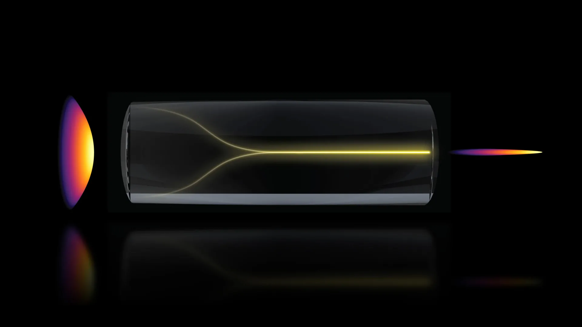
The genesis of this pivotal discovery stemmed from an unexpected observation that defied conventional expectations within the field of optics. The researchers had previously engineered a highly sophisticated device known as a "fiber shaper," designed to exert precise control over laser light as it propagates through a multimode optical fiber. These fibers are particularly advantageous for their capacity to transmit substantial amounts of optical power.
During their experiments, Cao incrementally elevated the laser’s power output to systematically probe the operational limits of the optical fiber. Contrary to the anticipated outcome, which would typically involve increased light scattering due to inherent microscopic imperfections within the fiber, a remarkable transformation occurred as the power approached a critical threshold. Instead of succumbing to disorder, the light abruptly concentrated itself into a single, exceptionally sharp, and well-defined beam.
"The inherent nature of these multimode fibers is characterized by a degree of intrinsic disorder," Professor You elaborated. "The conventional approaches to light manipulation required to overcome this disorder, especially when dealing with high power levels, have historically presented a significant and persistent challenge. However, through this self-organization phenomenon, we have achieved a stable, ultrafast pencil beam without the necessity of employing custom-designed beam-shaping optical components."
To consistently reproduce this remarkable self-organizing behavior, the research team meticulously identified two fundamental prerequisites. Firstly, the laser beam must be introduced into the optical fiber with an exceptionally precise alignment, entering at a zero-degree angle relative to the fiber’s axis, a requirement considerably more stringent than typically observed in standard optical setups. Secondly, the laser power must be elevated to a level where the light begins to exhibit direct interaction with the glass material constituting the optical fiber itself.
"At this specific critical power level, the nonlinear optical properties of the fiber material become influential enough to actively counteract the intrinsic disorder," explained Cao. "This dynamic interaction establishes a delicate equilibrium that effectively transforms the incoming, potentially scattered beam into a highly organized, pencil-shaped beam."
These specific experimental conditions are seldom explored by researchers because the prevailing practice is to meticulously avoid high power levels to prevent any potential damage to the delicate optical fibers. Moreover, the requirement for such precise alignment is not usually a primary concern, as multimode fibers are inherently designed to accommodate and transmit substantial quantities of optical energy. However, when these seemingly disparate factors are brought together under the precisely defined parameters of this study, the system demonstrates an extraordinary ability to generate a stable and coherent beam without the need for complex or specialized optical engineering.
"The elegance of this method lies in its simplicity," Professor You emphasized. "It is achievable with a standard optical setup and does not necessitate extensive specialized knowledge in the field."
Rigorous experimental evaluations have confirmed that the resultant pencil beam is not only exceptionally stable but also possesses an extraordinarily high level of detail when contrasted with beams generated through other conventional methods. Many existing laser beam configurations are prone to producing "sidelobes"—diffuse halos of light that invariably diminish the clarity and resolution of resulting images. In stark contrast, the beam produced by this novel technique remains exceptionally clean and tightly focused, minimizing these detrimental artifacts.
The researchers then proceeded to apply this advanced imaging methodology to visualize the human blood-brain barrier, a critically important cellular interface that serves as a protective shield for the brain against the ingress of potentially harmful substances, but which also presents a significant obstacle for the passage of many therapeutic drugs.
The ability of scientists to meticulously observe the movement of drug molecules through the vascular network of the blood-brain barrier and to confirm their successful penetration into brain tissue is paramount for the development of effective neurological treatments. Traditional optical imaging modalities typically operate by capturing sequential two-dimensional slices of the target area, necessitating multiple scans and considerable time to reconstruct a comprehensive three-dimensional representation.
By employing the newly developed pencil beam approach, the MIT team was able to generate rapid, high-precision three-dimensional images, simultaneously enabling the real-time tracking of protein uptake by individual cells.
"The pharmaceutical industry, in particular, has a vested interest in utilizing human-based tissue models to effectively screen drug candidates that can successfully traverse the blood-brain barrier, given that animal models often provide unreliable predictions of human responses," stated Professor Kamm. "The fact that this groundbreaking new method does not require cells to be genetically modified with fluorescent markers is a transformative development. For the first time, we are empowered to visualize the time-dependent entry of drugs into the brain and, crucially, to quantify the rate at which specific cell types internalize these compounds."
Dr. Spitz further highlighted the broader applicability of this technology: "Importantly, however, this approach is not confined solely to the study of the blood-brain barrier. It holds the potential to facilitate time-resolved tracking of a diverse array of compounds and molecular targets across various engineered tissue models, thereby establishing itself as a powerful instrument for the field of biological engineering."
The system successfully produced three-dimensional images at the cellular level, exhibiting demonstrably improved quality and achieving this feat at a speed approximately 25 times greater than existing methodologies.
"Typically, there exists a fundamental trade-off between image resolution and the depth of focus—researchers are limited in how deeply they can probe at any given time," Professor You explained. "However, with our innovative method, we have effectively overcome this limitation by generating a pencil beam that simultaneously offers both high spatial resolution and an extended depth of focus."
Looking towards the future, the research team intends to delve deeper into understanding the underlying physics governing the formation of this remarkable self-organizing beam and the precise mechanisms that drive its emergence. Their future research agenda also includes extending the application of this technique to other critical areas of biological inquiry, such as the imaging of neuronal activity, and actively pursuing avenues to translate this cutting-edge technology into practical, real-world clinical and research applications.
This significant scientific endeavor received crucial financial support from various sources, including MIT startup funds, the National Science Foundation (NSF), the Silicon Valley Community Foundation, the Diacomp Foundation, the Harvard Digestive Disease Core, a MathWorks Fellowship, and the Claude E. Shannon Award.
About the Author


