
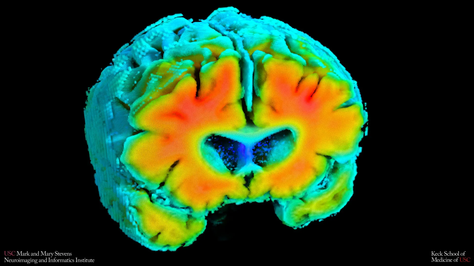
New investigations spearheaded by the Mark and Mary Stevens Neuroimaging and Informatics Institute (Stevens INI) at the Keck School of Medicine of USC suggest a profound connection between subtle alterations in the brain’s circulatory system and the nascent stages of Alzheimer’s disease. This groundbreaking research, published in the esteemed journal Alzheimer’s and Dementia: The Journal of the Alzheimer’s Association, postulates that the efficiency with which blood navigates the brain and the subsequent oxygenation of neural tissues may serve as an early, non-invasive marker for individuals at risk of developing this debilitating neurodegenerative condition.
The study’s methodology involved a comprehensive examination of older adults, encompassing both those exhibiting cognitive impairments and those functioning at a cognitively healthy baseline. Researchers meticulously assessed fundamental aspects of brain circulation and oxygen delivery through straightforward, non-invasive techniques. The findings revealed a statistically significant correlation: when the brain’s vascular network demonstrated characteristics akin to those observed in healthy aging, participants also exhibited fewer of the well-established pathological hallmarks of Alzheimer’s disease. These hallmarks include the accumulation of amyloid plaques, a key protein implicated in neuronal damage, and a reduction in the volume of the hippocampus, a brain region critically involved in memory formation and retrieval. This linkage implies that the integrity and functionality of the brain’s blood vessels might play a far more influential role in the initial phases of Alzheimer’s pathogenesis than previously emphasized, potentially offering a window for risk stratification before overt clinical symptoms manifest.
Amaryllis A. Tsiknia, the lead author of the research and a doctoral candidate at USC, emphasized that while amyloid and tau proteins have traditionally been considered the principal drivers of Alzheimer’s pathology, the role of blood flow and adequate oxygen supply to the brain is equally crucial. "Our findings underscore that a robust vascular system, functioning optimally, is intrinsically linked to enhanced cognitive resilience and healthier brain structures," Tsiknia stated, highlighting the interconnectedness of vascular health and neurological well-being. The study’s results suggest that the brain’s capacity to regulate blood flow and oxygen distribution in response to physiological demands, such as fluctuations in blood pressure and carbon dioxide levels, is a critical determinant of overall brain health.
To precisely quantify these intricate circulatory dynamics, the research team employed two sophisticated yet entirely painless diagnostic modalities. Transcranial Doppler (TCD) ultrasound was utilized to precisely measure the velocity of blood flow through the brain’s principal arterial pathways, offering a quantitative assessment of cerebral perfusion. Concurrently, near-infrared spectroscopy (NIRS) was employed to gauge the efficacy of oxygen diffusion from the blood into the cortical tissues located near the surface of the brain. These non-invasive approaches allowed for a detailed, real-time evaluation of the brain’s vascular performance without imposing any discomfort or risk upon the participants.
Following the acquisition of raw data from these instruments, the researchers applied advanced mathematical algorithms and computational modeling. This sophisticated analysis integrated the individual readings from TCD and NIRS into composite indicators of cerebrovascular function. These synthesized metrics provided a holistic picture of how effectively the brain’s circulatory system adapted to subtle physiological changes, reflecting its dynamic capacity to maintain optimal perfusion and oxygenation under varying conditions.
The study revealed a compelling pattern: individuals whose vascular health indicators more closely mirrored those of their cognitively unimpaired counterparts exhibited demonstrably lower levels of amyloid plaque accumulation and possessed larger hippocampal volumes. Both of these characteristics are recognized as protective factors against the development of Alzheimer’s disease, reinforcing the notion that compromised vascular function is not merely a co-occurring condition but may actively contribute to the disease’s progression.
Dr. Meredith N. Braskie, the senior author of the research and an assistant professor of neurology at Keck School of Medicine, commented on the significance of these vascular measures. "These vascular assessments are capturing a vital aspect of brain health," she explained. "They appear to align remarkably well with the findings obtained from magnetic resonance imaging (MRI) and positron emission tomography (PET) scans, which are the standard tools for investigating Alzheimer’s disease. This provides invaluable insights into the intricate relationship between vascular integrity and established biomarkers of Alzheimer’s risk."
Furthermore, the researchers observed a distinct trend among participants diagnosed with mild cognitive impairment (MCI) or dementia: they consistently demonstrated weaker cerebrovascular function when compared to the cognitively normal cohort. This finding provides substantial support for the evolving understanding of Alzheimer’s disease as a condition that encompasses not only the classic neurodegenerative processes but also significant contributions from vascular dysfunction, suggesting a continuum where declining vascular health is an integral component.
Dr. Arthur W. Toga, the director of Stevens INI, articulated the broader implications of this research. "These results bolster the accumulating evidence that Alzheimer’s disease is a multifaceted condition, involving substantial vascular contributions alongside the well-documented neurodegenerative changes," he stated. "By unraveling the complex interplay between blood flow regulation, oxygen delivery, amyloid pathology, and brain structural integrity, we are opening up promising new avenues for earlier detection and potentially for developing preventative strategies."
The potential utility of these non-invasive vascular assessment methods for broader screening is particularly noteworthy. When contrasted with MRI and PET imaging, TCD and NIRS offer considerable advantages in terms of cost-effectiveness and ease of administration. These techniques do not necessitate the injection of contrast agents, do not involve exposure to ionizing radiation, and do not require participants to undergo demanding cognitive tasks, making them significantly more accessible. This inherent simplicity could render them invaluable for large-scale population screening initiatives or for individuals who may be unable to tolerate or undergo more complex and resource-intensive neuroimaging procedures.
The authors of the study, however, prudently caution that the current findings represent a cross-sectional snapshot of the participants’ health and therefore cannot definitively establish a cause-and-effect relationship. To address this limitation and to explore the predictive power of these vascular markers, ongoing longitudinal studies are actively tracking the same cohort of participants. These long-term investigations aim to determine whether observed shifts in cerebrovascular function can reliably predict future cognitive decline or the efficacy of therapeutic interventions.
"Should we be able to effectively monitor these vascular signals over extended periods, we may gain the ability to identify individuals at heightened risk of Alzheimer’s disease at an earlier stage," Ms. Tsiknia elaborated. "This early identification could then pave the way for targeted interventions designed to improve vascular health, potentially slowing or even mitigating the progression of Alzheimer’s-related brain changes."
The research team responsible for this significant contribution includes Amaryllis A. Tsiknia, Meredith N. Braskie, Peter S. Conti, Rebecca J. Lepping, Brendan J. Kelley, Rong Zhang, Sandra A. Billinger, Helena C. Chui, and Vasilis Z. Marmarelis. This work was generously supported by funding from the Office of The Director, National Institutes of Health, under Award Number S10OD032285, and by the National Institute on Aging through grant R01AG058162.
About the Author


