
Major depressive disorder (MDD) stands as a pervasive global health challenge, contributing significantly to worldwide disability and imposing a considerable burden on individuals and healthcare systems. A substantial proportion of individuals diagnosed with depression, estimated at around 30%, develop what is termed treatment-resistant depression (TRD). This designation signifies a persistent struggle with the condition, where standard antidepressant medications fail to provide adequate symptomatic relief, leaving patients in a state of ongoing distress. In recent years, ketamine has emerged as a promising therapeutic agent, demonstrating a remarkable capacity for rapid antidepressant effects, particularly for those grappling with TRD. However, the precise neurobiological mechanisms underpinning ketamine’s swift efficacy in the human brain have remained an area of intense scientific inquiry, presenting a significant hurdle in the quest to optimize and individualize its clinical application.
Addressing this critical knowledge gap, a groundbreaking study, published on March 5, 2026, in the esteemed journal Molecular Psychiatry, has shed new light on ketamine’s intricate workings within the human brain. Spearheaded by Professor Takuya Takahashi from the Department of Physiology at Yokohama City University Graduate School of Medicine in Japan, the research team employed a sophisticated positron emission tomography (PET) imaging technique. This advanced methodology enabled them to directly observe and quantify changes in the density and distribution of glutamate $alpha$-amino-3-hydroxy-5-methyl-4-isoxazole propionic acid receptors (AMPARs) in the brains of individuals undergoing ketamine treatment. AMPARs are pivotal protein complexes that play a fundamental role in mediating synaptic transmission, influencing the efficacy of communication between neurons. Their function is intrinsically linked to synaptic plasticity – the brain’s ability to adapt and reorganize its connections – and the broader glutamatergic signaling pathways, which are profoundly affected by ketamine administration.
Professor Takahashi articulated the significance of their endeavor, stating, "Despite the recognized rapid antidepressant effects observed in patients suffering from treatment-resistant depression, the underlying molecular mechanisms operating within the human brain have eluded full comprehension." This research sought to bridge that chasm of understanding.
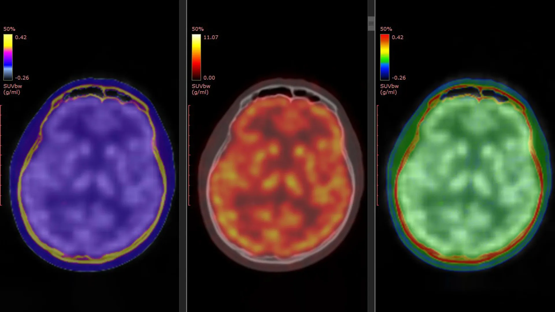
The cornerstone of this investigative effort was a meticulously developed PET tracer, designated as [$^11$C]K-2, previously engineered by Professor Takahashi’s laboratory. This specialized tracer possesses the unique capability to visualize AMPARs located on the surface of cells directly within the living human brain, a feat previously unattainable with such precision. Prior laboratory investigations and studies conducted on animal models had strongly implicated AMPAR activity as a key mediator of ketamine’s antidepressant properties. The present study, however, marks the first time direct, in vivo evidence has been gathered to confirm this hypothesis in human subjects.
To facilitate this crucial research, the investigators meticulously compiled and analyzed data derived from three distinct registered clinical trials conducted within Japan. The comprehensive study cohort comprised 34 patients formally diagnosed with treatment-resistant depression, alongside 49 healthy individuals who served as a control group, providing a vital baseline for comparison. Participants diagnosed with TRD were administered either intravenous ketamine or a placebo over a two-week therapeutic period. Crucially, PET brain imaging scans were conducted at two key junctures: prior to the commencement of treatment and again following the completion of the final infusion. This temporal imaging approach afforded researchers the unparalleled opportunity to meticulously track and compare alterations in AMPAR levels and their spatial distribution throughout the brain over the course of the intervention.
The analytical outcomes revealed a compelling pattern: individuals afflicted with TRD exhibited widespread deviations in AMPAR density when contrasted with their healthy counterparts. These observed anomalies were not uniformly distributed across the entire brain but were rather concentrated within specific neural circuits and regions. Intriguingly, the administration of ketamine did not induce a uniform alteration in AMPARs across all brain areas. Instead, the study established a significant correlation between improvements in depressive symptomatology and dynamic, region-specific modifications in AMPAR expression. Certain cortical regions demonstrated an augmentation in receptor density, suggesting enhanced glutamatergic signaling, while other areas, notably those implicated in reward processing, including the habenula, showed a discernible reduction in AMPAR levels. These intricate, location-dependent shifts in receptor distribution were found to be powerfully predictive of the degree of symptom amelioration experienced by the patients.
Professor Takahashi elaborated on these findings, emphasizing, "The antidepressant efficacy of ketamine in patients with treatment-resistant depression is facilitated by dynamic alterations in AMPARs within the living human brain." He further highlighted the pivotal role of their novel PET tracer, stating, "Employing the innovative PET tracer, [$^11$C]K-2, we were able to visualize how ketamine orchestrates changes in AMPAR distribution across specific brain regions and, critically, how these neurological adjustments correlate directly with improvements in the severity of depressive symptoms." These direct human observations provide robust empirical support for theoretical mechanisms previously elucidated through animal studies, firmly connecting them to tangible clinical benefits.
Beyond elucidating the fundamental neurobiological underpinnings of ketamine’s action, these findings hold significant potential for direct clinical application. The ability to visualize AMPAR density via PET imaging could evolve into a valuable biomarker. Such a biomarker could empower clinicians to more accurately assess and predict an individual patient’s likelihood of responding favorably to ketamine treatment, thereby enabling more informed therapeutic decision-making. Given the considerable challenges posed by the large percentage of patients who do not achieve remission with conventional antidepressant therapies, the identification of reliable biological indicators for predicting treatment success remains a paramount objective in the ongoing advancement of mental healthcare.
Ultimately, this pioneering research contributes substantially to the trajectory of developing more personalized and effective depression treatments. By providing a direct window into AMPAR activity within the living human brain, this study effectively bridges a longstanding divide between fundamental laboratory-based research and the practical realities of clinical psychiatry. The identification of AMPAR modulation as a central mechanism driving ketamine’s rapid antidepressant effects opens exciting avenues for future therapeutic strategies. The prospect of utilizing AMPAR PET imaging as a tool to guide and tailor treatment approaches offers the potential for a more individualized and precise therapeutic paradigm for individuals battling treatment-resistant depression. This comprehensive work promises to accelerate the development of highly targeted therapies, offering renewed hope and improved outcomes for those who have historically faced limited treatment options.
This research was generously supported by funding from the Ministry of Education, Culture, Sports, Science and Technology (through Special Coordination Funds for Promoting Science and Technology); the Japan Agency for Medical Research and Development (AMED), under grant numbers JP18dm0207023, JP19dm0207072, JP24wm0625304, JP25gm7010019, and JP20dm0107124; the Japan Society for the Promotion of Science KAKENHI, with grant numbers 22H03001, 20H00549, 20H05922, 23K10432, 19H03587, 20K20603, 22K15793, and 21K07508; the Takeda Science Foundation; the Keio Next-Generation Research Project Program; the SENSHIN Medical Research Foundation; and the Japan Research Foundation for Clinical Pharmacology.
About the Author


