
Groundbreaking research originating from Trinity College Dublin’s neuroscience department has revealed that infants merely two months old possess a remarkable capacity to discern and organize visual stimuli into distinct object classifications. This emergent ability surfaces significantly earlier than previously posited by the scientific community, indicating that fundamental underpinnings of perceptual processing are firmly established from the nascent stages of human life. The study, which employed a synergistic approach combining advanced brain imaging techniques with sophisticated artificial intelligence modeling, has illuminated the intricate cognitive processes at play in the minds of very young infants, offering unprecedented insights into their learning and understanding of the world long before they develop the capacity for language or intentional motor actions.
This pioneering work, published in the esteemed journal Nature Neuroscience, was conducted by a dedicated research consortium comprising members of the Trinity College Institute of Neuroscience (TCIN) and the School of Psychology. The collective effort aimed to address enduring questions about the internal world of infants and the nature of their visual experiences. Dr. Cliona O’Doherty, the principal investigator for this study who conducted the research while affiliated with Trinity’s Cusack Lab, articulated the significance of these findings, stating, "Parents and scientists alike have long pondered the internal landscape of an infant’s mind and the way they perceive their surroundings. This investigation underscores the profound richness of brain activity during the inaugural year of life." She further elaborated on the developmental implications, noting, "Despite the inherent limitations in communication due to a lack of language and refined motor skills at the two-month mark, infants’ minds were demonstrably not only forming representations of visual appearances but also actively engaged in the process of assigning these perceptions to specific categories. This substantiates that the foundational elements of visual cognition are operative from a remarkably early developmental stage, far sooner than anticipated."
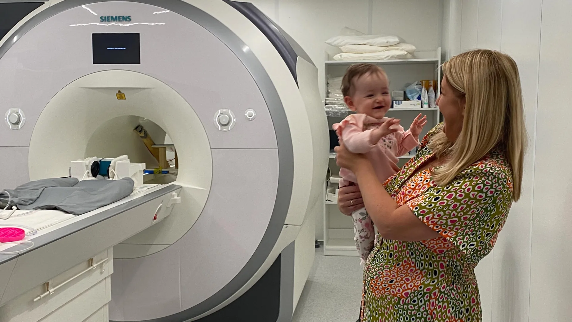
The investigative methodology involved the meticulous recruitment of 130 two-month-old infants through collaborations with the Coombe and Rotunda Hospitals in Dublin. Each participant was comfortably situated on a plush beanbag, equipped with noise-cancelling headphones, and presented with a curated selection of vibrant and engaging images designed to sustain their visual attention for periods of 15 to 20 minutes. This carefully controlled environment facilitated the utilization of functional magnetic resonance imaging (fMRI) to capture and record intricate patterns of neural activity as the infants observed images drawn from 12 familiar visual categories. These categories encompassed a diverse range of objects, including but not limited to felines, avian species, common playthings such as rubber ducks, everyday items like shopping carts, and natural elements like trees.
Following the acquisition of the brain scan data, the research team leveraged the analytical power of artificial intelligence models to meticulously decode how these varied visual categories were represented within the infant brain. By conducting a comparative analysis of neural activity patterns along the visual recognition pathways observed in both the AI models and the infants’ brains, the researchers were able to achieve a more profound understanding of the mechanisms underlying early perceptual categorization. Rhodri Cusack, the team’s leader and the Thomas Mitchell Professor of Cognitive Neuroscience at Trinity’s School of Psychology and TCIN, highlighted the study’s exceptional scope and potential impact: "This research represents the most extensive longitudinal investigation utilizing functional magnetic resonance imaging (fMRI) on awake infants to date. The wealth of data capturing brain activity unlocks entirely novel avenues for assessing the cognitive processes of infants at a very early age. Furthermore, it underscores the considerable potential of neuroimaging and computational modeling as diagnostic instruments for assessing very young children." Professor Cusack also expressed an optimistic outlook for future AI development, adding, "The learning capabilities of infants far surpass those of current AI models. By studying their accelerated learning processes, we aspire to catalyze the development of a new generation of AI systems that exhibit greater learning efficiency, thereby mitigating their associated economic and environmental costs."
The implications of these findings extend well beyond the confines of the laboratory, offering significant contributions to fields ranging from early childhood education to the advancement of artificial intelligence. Dr. Anna Truzzi, currently based at Queen’s University Belfast and a co-author of the published paper, emphasized the pivotal role of recent technological advancements in enabling this research: "Until quite recently, reliably measuring how specific regions of the infant brain processed visual information remained an insurmountable challenge. By integrating artificial intelligence with neuroimaging techniques, our study provides a truly unique perspective that significantly enhances our understanding of infant learning during their first year of life." She further elaborated on the critical developmental period, stating, "The initial year of life is characterized by exceptionally rapid and complex brain development. This study furnishes essential foundational knowledge that will be instrumental in shaping early-years educational strategies, informing clinical interventions for neurodevelopmental conditions, and fostering the development of AI approaches that are more closely aligned with biological principles."
Professor Eleanor Molloy, a neonatologist at Children’s Health Ireland and another co-author, underscored the broader societal importance of this research: "There is an urgent and pressing need for a more comprehensive understanding of how neurodevelopmental disorders impact early brain development, and awake fMRI technology presents a substantial opportunity to address this critical gap." Dr. O’Doherty is now continuing her research at Stanford University, while Dr. Anna Truzzi holds a position as Senior Lecturer in the School of Psychology at Queen’s University Belfast. The research was further complemented by artistic interpretations; artwork inspired by this groundbreaking research was created by artist Cian McLoughlin during his tenure as Artist in Residence at the Trinity College Institute of Neuroscience in 2024, alongside an accompanying exhibition essay.
About the Author


