
Groundbreaking new findings from a collaborative effort between neuroscientists at Trinity College Dublin and computational experts reveal that infants merely two months of age possess a sophisticated capacity for organizing the visual information they encounter into distinct object categories, a cognitive milestone previously thought to emerge much later in development. This research underscores the remarkably early establishment of fundamental perceptual building blocks within the infant brain, suggesting that a nascent understanding of the world is present from the very dawn of life.
By ingeniously integrating advanced neuroimaging techniques with cutting-edge artificial intelligence models, the research team has unveiled unprecedented insights into the intricate workings of infant cognition during their earliest months. These discoveries shed significant light on the neural processes at play long before the development of language or voluntary motor control, offering a profound glimpse into the formative stages of human perception and learning. The comprehensive study, detailing these remarkable findings, has been formally published in the esteemed scientific journal Nature Neuroscience, stemming from the collective endeavors of researchers affiliated with the Trinity College Institute of Neuroscience (TCIN) and the School of Psychology.
For generations, parents and scientists alike have harbored a deep curiosity about the internal world of infants, seeking to comprehend their subjective experience of the visual landscape. This pioneering investigation powerfully illustrates the inherent richness and complexity of brain function during the crucial first year of life. Dr. Cliona O’Doherty, the lead author of the study, who conducted this significant portion of the work while embedded within Trinity’s Cusack Lab, articulated the profound implications of the findings, noting that even at two months, when infants’ communicative repertoire is inherently limited by the absence of linguistic fluency and refined motor skills, their minds are actively engaged in not only processing the visual appearance of objects but also in assigning them to specific conceptual categories. This demonstrates that the foundational architecture for visual cognition is firmly in place at an astonishingly early stage.
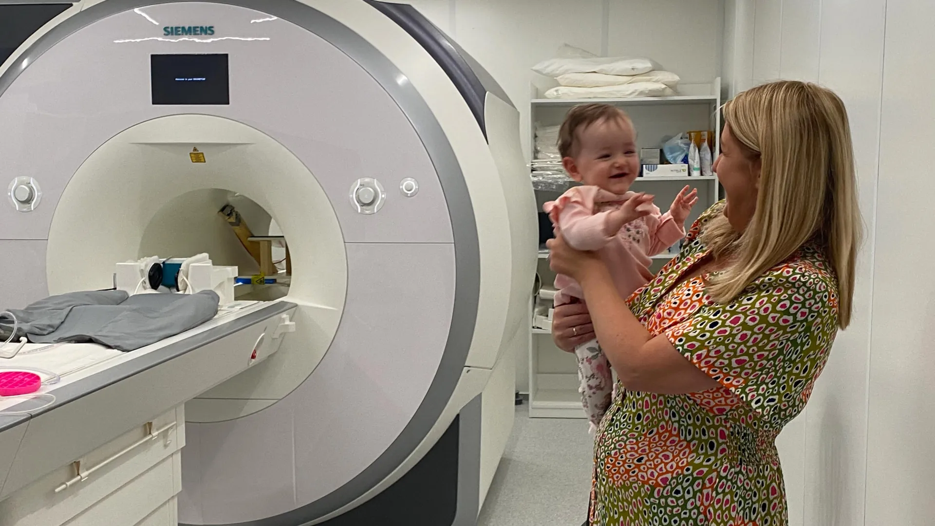
The experimental design involved the meticulous recruitment of 130 infants, all approximately two months old, through collaboration with Dublin’s Coombe and Rotunda Hospitals. Each participant was comfortably situated on a specialized soft beanbag, equipped with noise-canceling headphones, while being presented with a series of bright, engaging images specifically curated to sustain their attention for periods of 15 to 20 minutes. This carefully controlled environment enabled the researchers to employ functional magnetic resonance imaging (fMRI) to precisely record patterns of neural activity as the infants observed a diverse array of visual stimuli drawn from 12 familiar object categories, including common representations such as felines, avian creatures, bath toys, utility items like shopping carts, and natural elements like trees.
The subsequent analysis phase leveraged the power of artificial intelligence to meticulously decode the complex brain activity recorded during the imaging sessions. By training sophisticated AI models to recognize patterns associated with different visual categories and then comparing these algorithmic representations with the neural activity observed in the infant brains along established visual recognition pathways, the researchers were able to gain a granular understanding of how early object categorization operates. Rhodri Cusack, the project leader and Thomas Mitchell Professor of Cognitive Neuroscience at Trinity’s School of Psychology and TCIN, highlighted the study’s unique contribution as the largest longitudinal investigation utilizing fMRI in awake infants. He emphasized that the extensive dataset capturing neural activity offers a novel avenue for quantitatively assessing infant cognition at a very tender age and points to the significant potential of combining neuroimaging with computational modeling as a powerful diagnostic instrument for assessing very young children. Professor Cusack further noted that infants exhibit a learning capacity that far surpasses current AI systems, suggesting that studying their rapid and efficient learning processes could inspire a new generation of AI models, leading to more energy-efficient and environmentally sustainable artificial intelligence.
The broader significance of these findings extends well beyond the confines of the laboratory, offering crucial insights for various fields. Dr. Anna Truzzi, now a Senior Lecturer in the School of Psychology at Queen’s University Belfast and a co-author on the published paper, underscored the role of recent technological advancements in making this research feasible. She explained that until recently, reliably measuring how specific brain regions in infants process visual information was an insurmountable challenge. The synergistic combination of AI and neuroimaging employed in this study provides an exceptionally unique perspective, significantly deepening our understanding of infant learning throughout the first year of life. Dr. Truzzi stressed that the initial year of life is a period of extraordinarily rapid and complex brain development, and this research furnishes essential foundational knowledge that can inform early childhood education strategies, guide clinical interventions for neurodevelopmental conditions, and foster the development of AI systems that are more closely aligned with biological principles.
Adding to this perspective, Professor Eleanor Molloy, a neonatologist at Children’s Health Ireland and another co-author, pointed to the critical need for enhanced comprehension of how neurodevelopmental disorders impact early brain development. She asserted that awake fMRI technology holds substantial promise in addressing this imperative, offering a non-invasive and informative window into the developing infant brain. Dr. O’Doherty is currently based at Stanford University, continuing her research in neurodevelopment. The research was further complemented by artistic explorations; artist Cian McLoughlin, serving as Artist in Residence at the Trinity College Institute of Neuroscience in 2024, produced artwork inspired by this research, accompanied by an exhibition essay, further contextualizing the scientific discoveries.
About the Author


