
The period of adolescence represents a profound phase of transformation, extending far beyond observable physical and social maturation to encompass critical advancements in the brain’s intricate architecture. During these formative years, sophisticated cognitive functions, including strategic foresight, logical deduction, and the complex calculus of decision-making, undergo significant refinement and maturation. Despite the evident developmental progress, a comprehensive understanding of the precise mechanisms that sculpt the brain’s elaborate neural networks during this pivotal developmental epoch remains an ongoing scientific pursuit.
At the fundamental level of neural communication lie synapses, the crucial functional junctions that facilitate the transmission of information between neurons, forming the basis of all brain activity. For a considerable duration, the prevailing scientific paradigm posited a steady proliferation of synapses throughout childhood, followed by a pronounced reduction during adolescence. This established notion gave rise to the widely embraced theory of "synaptic pruning," a process wherein superfluous or inefficient neural connections are systematically eliminated. Consequently, it was hypothesized that an overabundance of this pruning mechanism could be implicated in the etiology of various neuropsychiatric conditions. Schizophrenia, a disorder characterized by profound disruptions in thought, perception, and emotion, including phenomena such as hallucinations and delusions, has frequently been associated with this theoretical framework of excessive synaptic loss.
However, recent investigations have begun to cast doubt upon this long-standing hypothesis. A distinguished cohort of researchers affiliated with Kyushu University has unveiled compelling evidence that challenges the prevailing view of adolescent brain development. Their groundbreaking study, disseminated in the esteemed journal Science Advances on January 14th, reveals a more nuanced developmental process than previously understood. The findings indicate that the adolescent brain is not merely engaged in the wholesale elimination of neural connections. Instead, it is actively engaged in the formation of novel, densely organized clusters of synapses within specific neuronal compartments during this critical developmental stage.
Professor Takeshi Imai, a senior researcher at Kyushu University’s Faculty of Medical Sciences, elaborated on the origins of this pivotal research. "Our initial objective was not to investigate neurological disorders," Professor Imai stated. "Following the development of a highly sophisticated tool for synaptic analysis in 2016, our curiosity led us to examine the cerebral cortex of mice. Beyond appreciating the inherent beauty of the neuronal structures, we were profoundly surprised to detect a previously unrecognized region exhibiting exceptionally high density of dendritic spines – the minute protrusions on dendrites where excitatory synapses are predominantly established."
The cerebral cortex, the outermost layer of the brain responsible for higher-level cognitive functions, is organized into six distinct layers, each contributing to the formation of remarkably complex neural circuits. Professor Imai and his team strategically directed their attention to the neurons residing within Layer 5. This specific layer plays a pivotal role in integrating information from a multitude of sources and subsequently transmitting outward signals, serving as the primary output pathway of the cortex. Given this central role, Layer 5 neurons function as critical control nodes in the brain’s sophisticated information processing pathways.
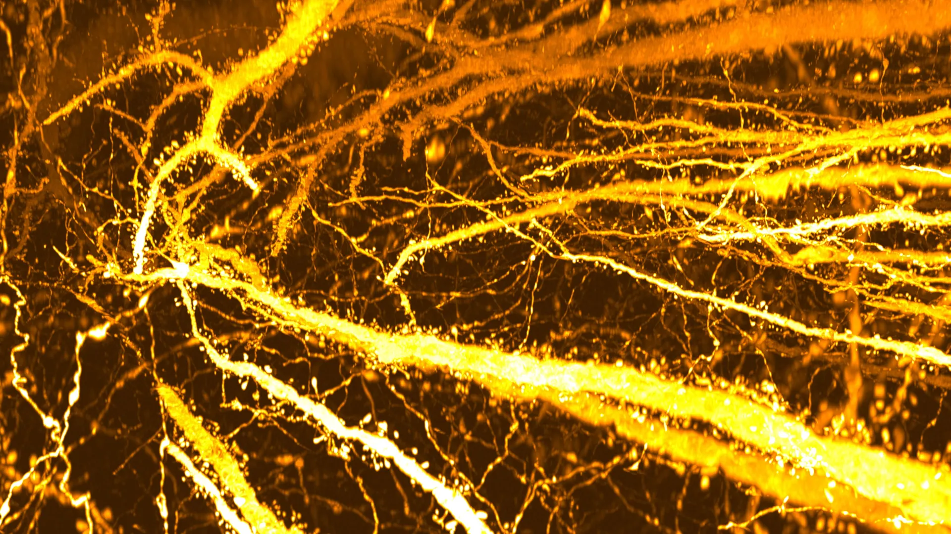
To facilitate an in-depth examination of these specialized cells, the research team employed a novel combination of techniques. They utilized SeeDB2, a specialized tissue clearing agent meticulously developed by Professor Imai’s laboratory, in conjunction with advanced super-resolution microscopy. This synergistic approach enabled the researchers to render brain tissue transparent, thereby allowing for the unprecedented mapping of dendritic spines across entire Layer 5 neurons with exceptional clarity and detail.
The meticulous mapping process yielded a remarkable and unexpected observation. A specific segment of the dendrite exhibited an unusually concentrated aggregation of dendritic spines, a phenomenon the researchers have aptly termed a "hotspot." Further rigorous analysis confirmed that this localized density of synapses is not present in the early stages of life but rather emerges specifically during the adolescent period.
To precisely delineate the temporal emergence of this developmental change, the researchers meticulously tracked the distribution of dendritic spines across various developmental milestones. In specimens of two-week-old mice, a stage preceding weaning, dendritic spines were observed to be distributed with relative uniformity across the neuronal structure. However, between the ages of three and eight weeks – a developmental window encompassing early childhood through adolescence – a striking increase in spine density was noted within a singular region of the apical dendrite. Over time, this localized proliferation of connections culminated in the formation of a distinct and densely packed synapse hotspot. "These findings strongly suggest that the long-accepted hypothesis of ‘adolescent synaptic pruning’ warrants a significant reevaluation," Professor Imai emphasized.
This newfound understanding of adolescent synaptic organization may also offer crucial insights into the developmental pathways of certain neurological disorders. Ryo Egashira, the study’s lead author and a former graduate student at Kyushu University’s Graduate School of Medical Sciences, posited a compelling link: "While synaptic pruning occurs broadly across dendrites, it appears that synapse formation also takes place in specific dendritic compartments during adolescent cortical development. A disruption in this precise formation process could be a pivotal factor in the manifestation of at least some forms of schizophrenia."
In an effort to empirically explore this hypothesis, the researchers examined mouse models engineered with genetic mutations known to be associated with schizophrenia, including alterations in the Setd1a, Hivep2, and Grin1 genes. These genetically modified mice exhibited typical early developmental trajectories, with normal spine density observed up to two to three weeks post-birth. However, a significant deficit in synapse formation became apparent during adolescence, critically impeding the proper development of the characteristic hotspot. This observation provides a potential alternative perspective to the long-held view of schizophrenia as primarily a condition characterized by excessive synapse loss, suggesting instead that difficulties in the constructive formation of new synapses during adolescence might play a central role. Nonetheless, the researchers prudently acknowledge that their study was exclusively conducted on murine models, and the direct applicability of these findings to primate or human brain development remains an open question requiring further investigation.
Looking toward the future, Professor Imai expressed his aspirations for continued research in this burgeoning field. "Moving forward, our primary goal is to precisely identify which specific brain regions are actively engaged in the formation of these novel synaptic connections during adolescence," he stated. "This will provide invaluable information regarding the actual neural circuits that are being constructed during this critical developmental window. A deeper understanding of the intricate ‘how’ and ‘when’ of these connections forming promises to significantly advance our knowledge of both fundamental brain development and the underlying mechanisms of neuropsychiatric disorders."
About the Author


